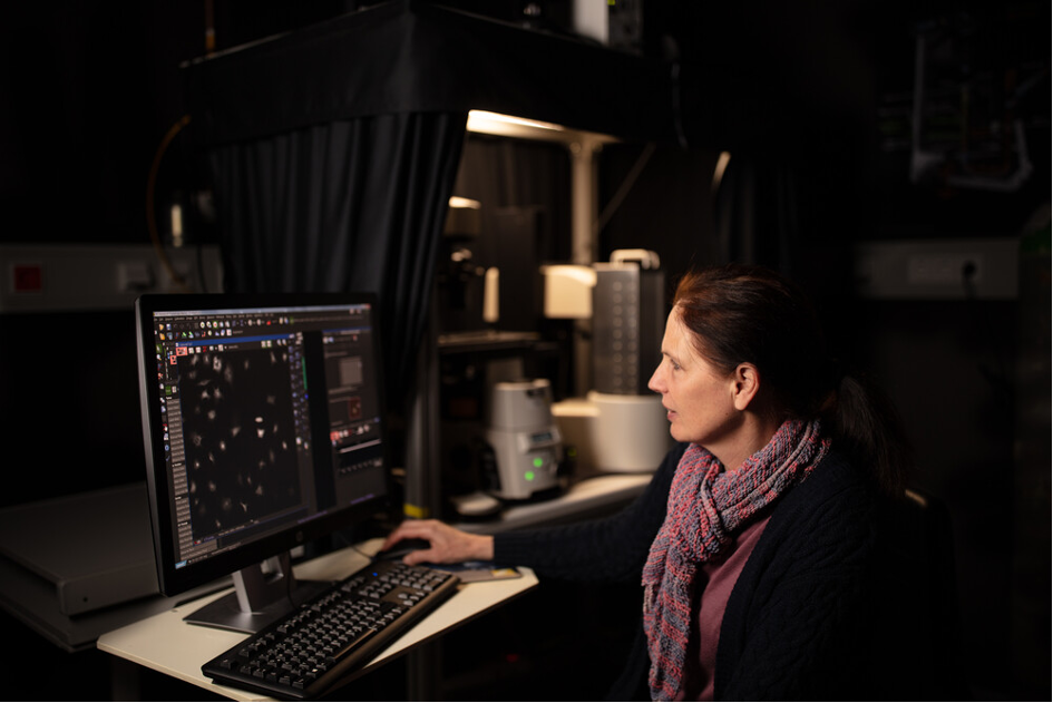
A global pandemic, climate change… the world is changing, and so is science. In this discussion, Beate Neumann, Senior Scientific Officer at EMBL’s Advanced Light Microscopy Facility (ALMF), part of the Euro-BioImaging’s EMBL Node, explains the ALMF approach to user projects in this new context. Working hand-in-virtual hand with her users, she runs better, more efficient experiments for the benefit of science – and the planet.
In April 2020, when the first global lockdown due to COVID-19 hit, running an experiment remotely was almost unthinkable for Beate Neumann, who has coordinated microscopy based high-throughput screening with international and national users at EMBL for many years. “User projects usually involve several visits to our facility,” says Beate. “These visits are important to get to know the user and for on-site training before the experiment actually starts. Using many different devices – none of which are familiar to our users - is quite challenging. It is my job to help users with their project from the beginning to the end of their experiment.”
But since 2020, this approach has been evolving. Facility coordinators are integrating virtual work practices into their interactions with users – and discovering that they work. “We are able to do excellent science – while at the same time reducing our carbon footprint,” says Beate. In this article, Beate Neumann shares some experience for a successful high-throughput screening project in the new global context.
Step 1: “Virtual” consultation & evaluation of the samples/cell line
For high-throughput screening projects, the first contact between a potential user and the facility staff is a consultation phase. “It is very important to discuss the biological question the user wants to address. Facility staff have so much experience, we can easily recommend a technology based on what they want to do. Sometimes a user thinks they want to do an experiment with high-throughput project on a genome-scale, but after consulting with them, they realize that only smaller scale projects are feasible due to time and costs. In addition, the sample read-out is key in order to answer the biological question. Therefore, we need to decide which image resolution, image analysis, and throughput is necessary for a successful project while at the same time keeping an eye on time and available funds for the project.”
Over 10 years ago, Beate Neumann had already transformed the consultation phase with the user into a virtual process. It can take place via Skype or Zoom - and it’s just as effective as an in-person consultation.
“We can get a really good idea of what the user wants to do remotely. We discuss with them, we read their scientific references. There’s really no need to travel,” says Beate.
But what about the user’s cell line? Typically, once the biological question has been clarified, the facility coordinator must evaluate the samples / cell lines – to make a recommendation as to what type of imaging approach should be used.
“Evaluating the samples/ cell lines is crucial to make an experiment as efficient as possible,” says Beate. “But this also can be done remotely. The user simply sends us raw images, of e.g. treated and untreated cells. Once we have the image file, we can consult with Christian Tischer, Imaging analysis expert running at EMBL Heidelberg the Centre for Bioimage Data Analysis in close collaboration with the ALMF, who will support the user in image analysis of the high-throughput data. He is able to tell us if the read-out is good enough to answer the biological question.”
Once the cell line has been evaluated, if both sides agree on how to continue, then the project moves on to Step 2.
Step 2: Setting up the control assay & virtual microscopy
Now is the time to set up control experiments – and discuss the image analysis pipeline. Here, Beate works very closely with the user - remotely. The user’s biological question may be refined as the scale of the experiment and the screening format are determined. In parallel, the user will start to familiarize themselves with the open source image analysis software used at EMBL.
Once everything is clear, the user performs control experiments at their home institutes using the future imaging plates - or if they prefer, they can send their samples to EMBL for testing and/or preparation. siRNA coated plates ship really well at room temperature, so this is not a hurdle. For testing, the user joins Beate at the microscope via a remote connection from their home institute. The user sees everything as if they were there - and can remotely adjust the microscope settings.
“Prior to corona, we did the control assay often in person. But now we do them remotely – which is much better for the environment.”
At this point, Beate is able to give the user a cost estimate, which provides the basis for the budget they will request in their grant.
Step 3: The experiment is underway
In the last phase of the project, the user travels to EMBL to carry out their experiment.
Once the user is on-site, they undergo training for all of the equipment, not just the microscopes. They are also trained in cell seeding, coating, image analysis… And then, the hard work begins. A relentless job of preparing and screening plates day in and day out that can last for several weeks.
Prior to the global pandemic, users came to EMBL 2-3 times over the course of an experiment. With the new work practices, travel obligations are drastically reduced – saving time, money - and the planet. Nevertheless, running a high-throughput experiment is a long process, and users have ample time to network on-site – an important facet of scientific collaboration.
In this new world, Beate and the ALMF team at EMBL are as present as ever, helping their users every virtual step of the way – from preparing the plates to running the experiment to image analysis. And many rich collaborations are underway - with very promising scientific outcomes.
Remote microscopy & training at Euro-BioImaging Nodes
Many of our Nodes are implementing remote access workflows in response to the global pandemic. Here is a selection of articles describing new practices from remote training to remote microscopy. If you have a story that you want to share, please contact us at info@eurobioimaging.eu.
The Euro-BioImaging EMBL-Node offers a collection of state-of-the-art microscopy equipment and image processing tools. This Node supports in-house scientists and visitors in using microscopy methods for their research and regularly organizes international training courses to teach basic and advanced microscopy methods. The services provided include project planning, sample preparation, microscope selection and use, image processing and visualization. Through Euro-BioImaging, life scientists, regardless of their affiliation or area of expertise, can apply to access services from 33 Nodes, composed of internationally-renowned imaging facilities.
More news from Euro-BioImaging
July 28, 2026
Global BioImaging Advance the Conversation on Sustainable Biomedical Imaging Facilities
How can biomedical imaging facilities remain scientifically excellent while becoming more financially, environmentally, and operationally sustainable? These questions were at the heart of…
Moara Lemos: New insights from Global BioImaging’s Job Shadowing programme
Moara Lemos is a Brazilian researcher and imaging scientist. Her award winning research pushes the boundaries of structural biology, using advanced cryo-electron microscopy…
A Multiplexed Vision: Highlights from the Special Edition Virtual Pub on Spatial Transcriptomics
On June 12th, Euro-BioImaging hosted its latest Special Edition Virtual Pub, bringing together over 200 researchers and technology developers working at the cutting…