Understanding the effects of ionizing radiation on developing organisms is a critical area of research in radiobiology and developmental biology. Embryonic tissues are particularly sensitive to radiation due to their rapid cell division and differentiation, making them a valuable model for studying early-stage damage and repair mechanisms.
Prof. Roberto Pacelli, a radiation oncologist from the University of Naples, applied for access at the Molecular Imaging Italian Node for investigating the effects of ionizing radiation on early embryonic development in chicken embryos, with a particular focus on brain damages, combining experimental irradiation, advanced imaging, and histological analysis.
Roberto Pacelli is full professor of Imaging and Radiotherapy at the University Federico II of Napoli, and Head of Radiation Oncology and Radiosurgery Department of the University Hospital. His research interests are related to the optimization of cancer therapy through better comprehension of the radiobiology of cancer and normal tissues and of the interaction between drugs and ionizing radiation. He applied to the SEELIFE funding, granted by the Italian Ministry of Research for strengthening the Italian Euro-BioImaging network, with a project on the evaluation of early damage induced by radiation therapy on embryos.
A non-invasive technique
The chicken embryo is widely used in developmental studies because it allows for controlled manipulation and observation of embryogenesis within a defined time window. On the other hand, recent advances in non-invasive imaging techniques, such as magnetic resonance imaging (MRI), provide new opportunities to monitor structural and functional changes in vivo without disrupting development. For this reason, Prof. Pacelli and the CNR Institute of Biostructures and BioImages in Naples, part of our Molecular Imaging Italian Node, designed an imaging study to follow up chicken embryos exposed to radiation via MRI.
“Access to the Euro-BioImaging facility had a significant positive impact on the study, enabling high-quality data acquisition and supporting the successful completion of the project.”
- Prof. Roberto Pacelli, University of Naples
For this study, fertilized chicken eggs were initially incubated under controlled conditions to support normal growth. At a specific developmental stage, a group of embryos was exposed to a planned dose of radiation, while a control group underwent the same handling without irradiation. Embryonic development was monitored through high-field MRI at several time points, allowing to observe structural and developmental changes non-invasively. The acquisition protocol comprised whole-egg high resolution T2 weighted sequences and brain diffusion tensor imaging. On day 15 of development, all embryos underwent histopathological assessment, enabling direct comparison between imaging findings and microscopic tissue analysis.
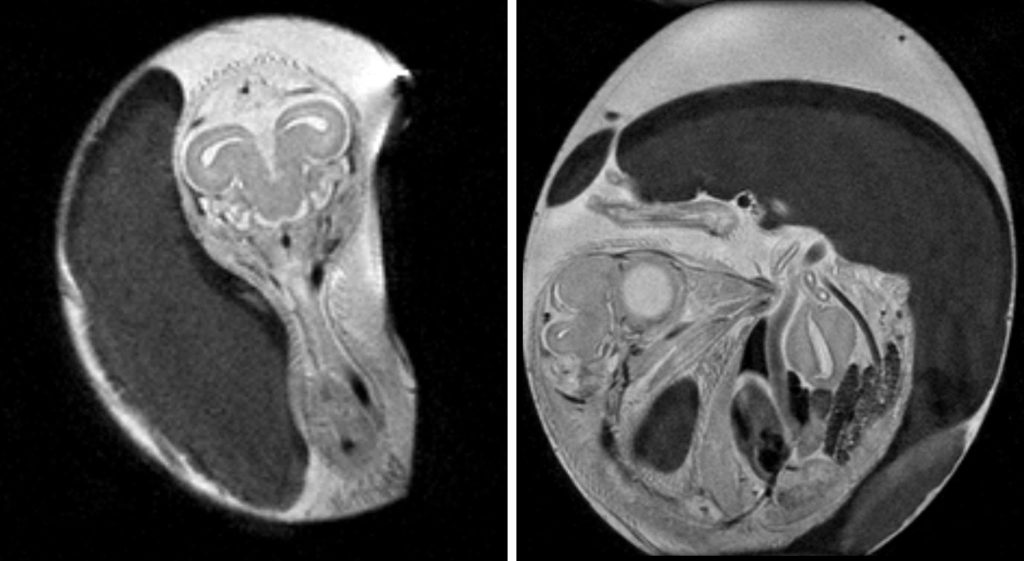
High-resolution T2-weighted MR images of chicken embryo in coronal (left) and sagittal (right) direction. Image courtesy of Prof. Pacelli.
Understanding how radiation affects organogenesis
Combining advanced imaging with histological analysis in a controlled embryonic model, this study provided a unique opportunity to explore how ionizing radiation affects organogenesis, especially brain development, in a way that is directly relevant to understanding long-term tissue sensitivity and damage. The study yielded promising results, establishing for the first time a quantitative MRI framework for radiobiological investigations in the in ovo chicken embryo model, with significant structural abnormalities or growth delays observed in irradiated samples.
The insights gained within this study will help inform safer clinical practices and contribute to translational research in radiation protection, pediatric oncology, and normal tissue preservation.
More news from Euro-BioImaging
July 20, 2026
Strengthening research-industry partnerships: results from the EVOLVE industry job shadowing pilot
The EVOLVE project, supported by European Union funding, recently completed a pilot programme designed to deepen the relationships between academic research infrastructures…
Imaging-Driven Neuroscience, Responsible Research, and the Power of Advocacy: Highlights from FENS Forum 2026
Euro-BioImaging was pleased to participate in the FENS Forum 2026, Europe’s largest international gathering dedicated to advancing neuroscience (>8000 participants). Throughout…
Euro-BioImaging Highlights the Value of Research Infrastructures at the FENS 2026 Satellite Symposium hosted by EBRAINS
Euro-BioImaging was pleased to participate #FENS2026 Satellite Symposium “Accelerating Your Neuroscience Research through the European Research Infrastructures” organised by EBRAINS, to learn…