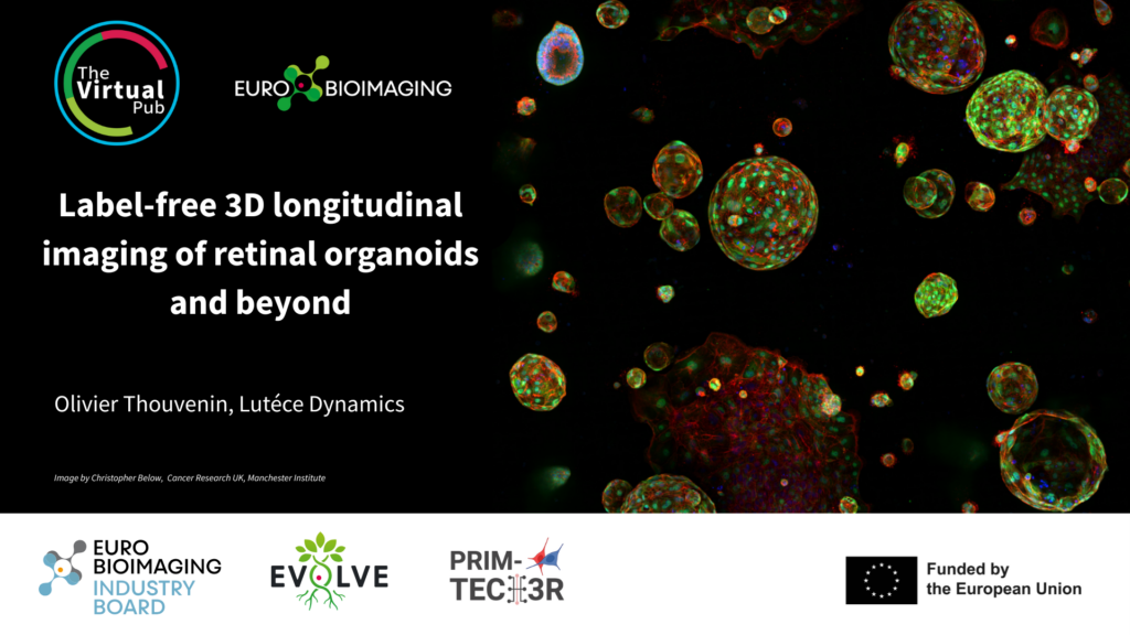
We will host the Special Edition Virtual Pub "Imaging Organoids" on Friday, January 30, from 1-3 pm CET. It will focus on methods used to image organoid systems. As part of this event, Olivier Thouvenin, Lutèce Dynamics, will give a talk entitled "Label-free 3D longitudinal imaging of retinal organoids and beyond" (description below). Join us to hear this and other exciting presentations introducing a range of methods used to image organoid systems - from lung buds to mini-brains.
Title: Label-free 3D longitudinal imaging of retinal organoids and beyond
Author: Olivier Thouvenin, Lutèce Dynamics
We have developed a new imaging modality that enables label-free and non-destructive imaging of thick biological samples, and in particular organoids. Our technology is based on interferometric microscopy, named dynamic full field optical coherence tomography, which allows 3D detection of endogenous optical contrasts linked with refractive index differences. By quantifying the biological movements of organelles detected with our microscope, we were able to demonstrate that this technology can detect all living cells in biological tissues and characterise how the cells transport their internal material, which is strongly correlated with their metabolism. In collaboration with the Vision Institute, we were able to study the growth of the same retinal organoid over several weeks and characterise the degeneration of retinal organoids derived from patients over several months.
Lutèce Dynamics is a one year-old start-up aiming to disseminate and commercialize this technology, which will empower life science researchers with live non-destructive evaluation of their samples, from 2D and 3D cell cultures to explants and small animals. We are commercializing the VertXTM, a small module (25x70x30 cm3) that plugs into the back of any commercial microscope and can be coupled with any other imaging technology. It allows us to characterise the living cells of an organoid to a depth of several hundred micrometres, at resolutions of around 0.3x0.3x0.5 um3, and over fields of view of several mm².
During this presentation, I will briefly detail the technical principles of dynamic full field OCT, and will give several examples of biological applications enabled by our module. We hope to explore new applications and to find partners within the Euro-Bioimaging community to test and disseminate our technology on a larger scale!
More news from Euro-BioImaging
October 7, 2026
French volume EM community comes together for its first scientific meeting
On 28–29 September 2026, the growing French volume electron microscopy (vEM) community came together in Bordeaux for the first France VolumeEM Scientific Days.
Euro-BioImaging contributes life science perspective to future European Research Infrastructure frameworks
Euro-BioImaging is contributing its experience as a distributed life science Research Infrastructure to European discussions on the future frameworks for Research Infrastructure access,…