Laser Microdissection: Extract specific regions from your sample
Published March 9, 2023

Country
Node
Category
Want to look at tumor vs. healthy tissue within a sample? Laser microdissection enables you to select a material based on its spatial or qualitative traits. We spoke to Natalia Nowak, a biologist at the Laboratory of Imaging Tissue Structure and Function - light microscopy core-facility of Nencki Institute of Experimental Biology in Warsaw, Poland, part of our Polish Advanced Light Microscopy Node. Her facility has recently added this exciting new technique to their portfolio and is now accepting applications for microdissection projects as part of the Euro-BioImaging Proof-of-Concept study. Read more about this technology and what it can be used for in the interview below.
My name is Natalia Nowak. I work as a biologist in the Laboratory of Imaging Tissue Structure and Function - light microscopy core-facility of Nencki Institute of Experimental Biology in Warsaw, Poland. My facility is part of the Polish Advanced Light Microscopy Node. For many years, our facility has offered a variety of fluorescent wide-field and confocal microscopes for live and fixed samples imaging, and Laser Microdissection (LMD) is an exciting, new technique we recently added to our portfolio.
We are today talking about Microdissection imaging. Please provide a short summary of this type of imaging and list some applications:
Laser microdissection (LMD) is a technique which allows users to selectively cut out regions of interest from a sample using a high-power laser. The extracted regions can then be collected specifically and subjected to further experiments, which may involve DNA, RNA and proteomic analysis. Therefore, LMD enables researchers to select the material for sequencing, proteomics or other analysis methods based on its spatial or qualitative traits: tumour vs healthy tissue, cells expressing fluorescent markers or specific proteins detected with antibodies etc. This allows normally bulk analysis methods, such as sequencing, to be specifically applied to selective parts of a sample with cellular and even sub-cellular levels of precision.
The sample is usually a fixed tissue slice, but microdissection of live cells and their further culturing is also possible. Using different objectives available on our system we are able to cut out tissue fragments, single cells and even single chromosomes. The cut regions fall gravitationally into the collector module below the sample and can be sorted into specific tubes or wells based on the configured settings. This means that users can collect many individual samples for further analysis from a single tissue slice.
Tell us a bit more about a specific project that was done in your facility using this technology? What scientific questions were you addressing?
As we have included laser microdissection in the facility offer just recently, there is only one ongoing research project which uses this method. The experiments’ goal is the analysis of the proteome of different neuronal populations in the spinal cord.
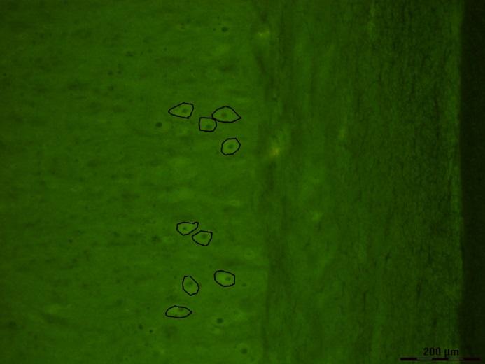
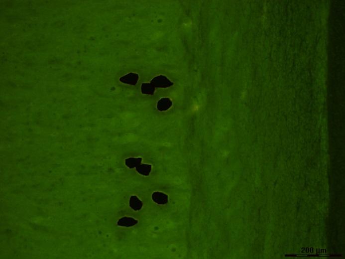
Spinal cord slice with fluorescently labelled cells marked for dissection (left) and after dissection (right). Authors: Olga Gajewska and Natalia Nowak, Nencki Institute of Experimental Biology.
What are some advantages of this technique that make it suited to addressing this type of question?
The technique allows to selectively extract only the cells of interest which are rarely found in the tissue and comprise only a small portion of whole-sample proteins or nucleic acids. This means that further analysis techniques can be used much more precisely and effectively and reveal variations of e.g. protein expressions in specific cell populations that would be hidden when looking at overall protein expression levels in the tissue.
The system available in our facility offers both transmitted-light and fluorescence imaging, therefore, the processed tissues can be stained with a variety of dyes: immunohistochemical or fluorescent to distinguish between different cellular populations.
What other services do you provide in your facility that would be useful in combination with this type of imaging?
Our team can advise you on tissue staining and preparation of the specimen, help you choose the best microscope and, of course, provide you with assistance when using the systems. Specifically, before performing laser microdissection, you can image your specimen on some other systems available in our facility to make sure the sample and staining are satisfactory. Our automated, wide-field fluorescent slide scanner would be especially useful in reviewing and assessing multiple samples before proceeding with microdissection to make sure you will get the best results in the shortest time possible.
How to apply:
Laser microdissection is part of the Euro-BioImaging Proof-of-Concept study. The Proof-of-Concept study makes it possible to introduce exciting, new imaging technologies to our portfolio that were previously unavailable via our network. We are currently accepting applications to use these technologies at participating Nodes as part of the Proof-of-Concept study. Be part of this study - and contribute to community-wide continuous technological innovation!
All scientists, regardless of their affiliation, area of expertise or field of activity can benefit from Euro-BioImaging’s pan-European open access services. Potential users of these new technologies are encouraged to submit project proposals via our website. To do so, you can log in to access our application platform, choose the technology you want to use and the facility you wish to visit, then submit your proposal. All applications will be processed by the Euro-BioImaging Hub. As usual, users will benefit from advice and guidance by technical experts working at the Nodes, training opportunities, and data management services.
For more information: info@eurobioimaging.eu
Pictures:


More news from Euro-BioImaging