
March 30, 2026
Euro-BioImaging at the EOSC ESFRI meeting in Milan
Euro-BioImaging was delighted to attend the ESFRI/EOSC Policy Workshop on “EOSC and Research Infrastructures: Opportunities and Strategies” in Milan from March 15-16, represented by…

It all started at the Molecular and Biophysical Bases of Photosynthesis Conference held in Venice in May 2022. Sara Natale, then a PhD researcher at the University of Trieste, got a helpful tip from another conference attendee about FLIM, an imaging technique that could support her research into stem photosynthesis in woody species. The conference attendee also put her into contact with Prof. Herbert van Amerongen at the Euro-BioImaging WISH Node in Wagenigen, a Node that offers open access to advanced imaging instrumentation developed for the particular purpose of studying photosynthesis in vivo. Thanks to the Euro-BioImaging Italian Fund for User Access, Sara Natale was able to visit the Node in Wagenigen and observe chloroplasts involved in stem photosynthesis in Fraxinus ornus with FLIM.
Photosynthesis, the process by which plants convert light into sugar and oxygen, occurs not only in leaves but also in organs optimized for other functions, such as stems for certain plants. Recent literature suggests that stem photosynthesis can help plants to cope with detrimental impact of drought. However, knowledge about stem photosynthesis is still very poor. The objective of Sara Natale’s PhD research was to characterize differences in chloroplasts in the bark & wood as well as identify/compare photosynthesis systems in the stem of different years of Fraxinus ornus.
Understanding drought resistance
“The hypothesis of my PhD research was that stem photosynthesis might be an ally for trees/plants facing drought,” explains Sara. “I started my PhD by applying optical methods such as spectral reflectance, fluorescence microscopy, chlorophyll fluorescence imaging, and in vivo spectroscopy to study both structural and functional characteristics of bark and wood chloroplasts as well as in the corresponding leaves, considered as reference controls. By studying these systems, I hope to provide a comprehensive characterization of bark and wood chloroplasts in woody plants in order to better understand how stem photosynthesis can be involved in tree resistance/resilience to drought stress.”
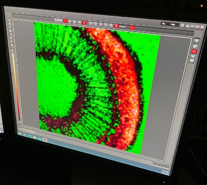
When Sara learned about Fluorescence Lifetime Imaging Microscopy (FLIM) at a conference, she was intrigued. FLIM is a widely used imaging technique, which allows mapping of fluorescence lifetimes with (sub)nanosecond time resolution and a diffraction-limited spatial resolution of approximately 250 nm. An advantage of this method is that allows to determine reliable distributions and properties of Photosystem I (PSI) and Photosystem II (PSII). Indeed, one can directly quantify the photosystems characteristics in the ex vivo samples, while with other methods one needs to isolate pigment-protein complexes or work with low-temperature (77K) fluorescence, and these methods are potentially prone to artifacts. But the trip to Wagenigen would be expensive and there was no budget at her home institute for her undertake it.
Enter the Euro-BioImaging Italian Fund, an initiative from the Italian Ministry of Universities and Research, to support Italian scientists visiting Euro-BioImaging Nodes across Europe. The Fund also incites researchers from around the world to visit Euro-BioImaging Nodes in Italy. Advised by Prof. Herbert van Amerongen, Sara applied for this funding and got it.
To carry out her project, Sara travelled to Wagenigen by train. She booked a temporary accommodation for her stay – it would take two full weeks for her to complete her project.
Project support from the experts
“This was a great experience for me. Prof. Herbert van Amerongen and his Phd student, Ludovico Caracciolo, both helped me a lot, from teaching me how to use the microscope to analyzing my data. With their help, I also applied a methodology for leaf photosynthesis developed in the WISH Lab and compared the stem and leaf photosynthesis processes.”
“On top of this, I gained a new skill. I would now know how to use FLIM independently and identify which data was accurate enough to take into consideration.”
“Using FLIM really helped me to answer my research question in a very precise way. And the high quality results I gained would not have been possible without this collaboration. As a result, I am writing paper,” concluded Sara.
Sara Natale now lives in Padua where she is doing a post-doc for the next 2 years. Her next projects will examine herbaceous and woody plants and she is considering the use of Environmental Scanning Electron Microscope (ESEM), Scanning Electron Microscope (SEM) or Transmission Electron Microscope (TEM) to gain insight into her samples.
March 30, 2026
Euro-BioImaging was delighted to attend the ESFRI/EOSC Policy Workshop on “EOSC and Research Infrastructures: Opportunities and Strategies” in Milan from March 15-16, represented by…
March 30, 2026
Euro-BioImaging was delighted to attend the Public Awareness & Engagement of Research Infrastructures (PAERI) conference, represented by External Communications Officer, Marianna Childress-Poli. This year’s…

March 26, 2026
The Madrid Advanced Microscopy Center (MAdMiC) is the first Euro-BioImaging Node in Madrid (Spain). It is formed through the collaboration and close work of…