

July 9, 2026
Euro-BioImaging Visits the NORMOLIM Node at the 2026 180°N Conference
In April 2026, the Med-Hub Head of Operations Alessandra Viale attended the 2026 180°N Conference, held at the beautiful Nye Hjorten Teater…
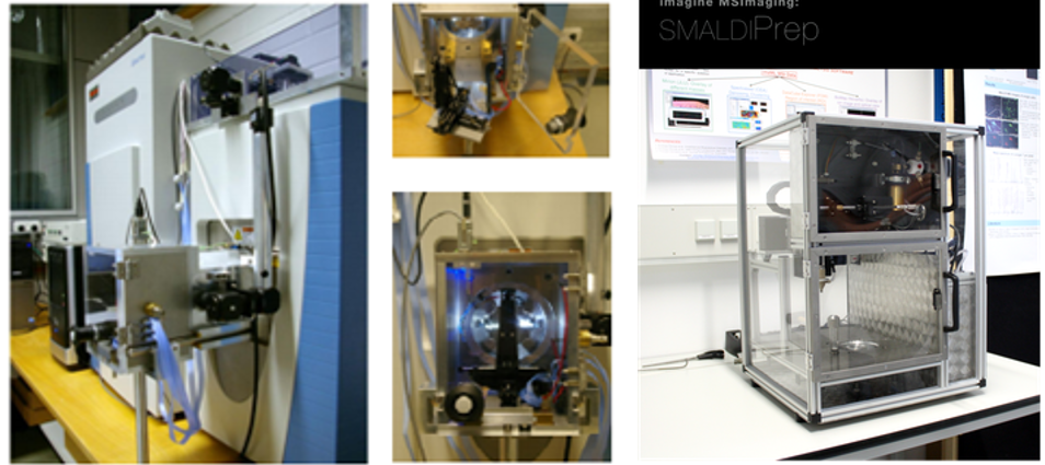
Let me tell you about Mass-Spectrometry-based Imaging (MSI) at the Bioimaging facility, IBBC-CNR. Our technology is based on an AP-SMALDI10 mass spectrometer with a dedicated home-built matrix pneumatic sprayer (SMALDI prep) and software for images generation (Mirion). This particular instrument couples an orbital trapping mass spectrometer (QExactive, Thermo Fisher) with an atmospheric-pressure scanning-microprobe MALDI imaging source (AP-SMALDI10, TransMIT) that is equipped with a focused laser beam able to scan the sample with a 7µm lateral resolution.
Mass Spectrometry Imaging (MSI) is a technique that uses mass spectrometry to visualize and localize analytes of interest by analysing their molecular masses without the need for labelling, without any a priori knowledge of the sample and without altering the morphology of the tissue. With MSI it is possible to correlate the information on the chemical composition of a biological sample with the morphology of the sample. The analysis consists of collecting mass spectrum spot by spot across the sample until the entire sample is scanned. By choosing a peak in the resulting spectra that corresponds to the compound of interest, the MS data is used to identify and check its distribution all over the sample. In this way, it is possible to obtain pictures of the spatially resolved distribution of the molecule of interest pixel by pixel. The power of MSI is that it enables us to perform studies of metabolomics, proteomics and lipidomics without needing to tag the molecules of interest. Some specific applications include the analysis of metabolite or lipid distribution, distribution of small peptides and glycan profiling across tissue sections.
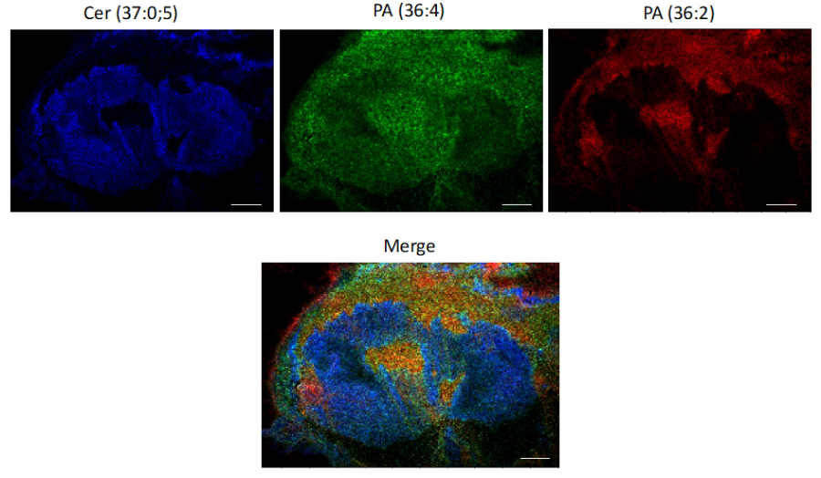
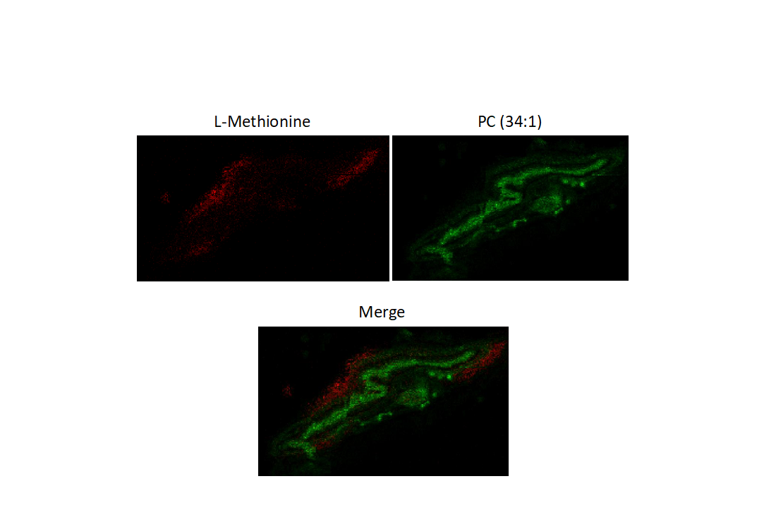
A recent project carried out in our facility using MSI was the analysis of the biodistribution and metabolism of L-Methionine in retinal tissue from rodents following the study of the spatial distribution of lipids that compose the retinal sections. We focused on analysis of the distribution, levels, and metabolic conversion of the exogenously administered 15N-L-Methionine in the eyes of the rodent.
Images of lipid distribution:
Images of L-Methionine distribution:
Localization of metabolites by conventional imaging techniques requires the addition of a tag that can subsequently be visualized by fluorescence or other imaging modes. While these conventional imaging modes have superior resolution, the addition of a tag, especially on a small metabolite, can affect the structure and behavior of the tagged molecule and thus cannot guarantee that the tagged metabolite actually follows physiologically relevant pathways. On the contrary, MSI can provide spatial information using untagged metabolites and thus provides a more physiologically relevant localization of information compared to other techniques.
Sample preparation, sample treatment and sample storage are critical issues in MSI. A careful sample preparation is necessary to prevent the possibility of delocalization of analyte(s). The choice of the correct matrix that is applied to the sectioned samples is also crucial.
In addition to MSI, our facility provides access to other modes of imaging inlcuding fluroescence and light microscopy, electron microscopy and Raman microscopy. Thus, our facility presents a unique opportunity for correlative imaging where MSI can be coupled to other modes of imaging to provide enhanced information.
MSI is an emerging technology and is not widely available. The technology is available only at a few places in Europe, and in Italy there are only two labs that have this instrument. Further, as mentioned earlier, our facility, unlike many others where the technique is available, provides also other imaging techniques in parallel which helps in developing correlative imaging modes. And on a lighter note, when you visit us, you can also enjoy the best pizzas in Naples!
In July 2020, Euro-BioImaging launched a Proof-of-Concept study - in collaboration with its Nodes - making it possible to access six previously unavailable imaging technologies. This article – the fourth in a series - features interviews with Euro-BioImaging Node staff, shedding light on some potential applications of these technologies - and providing advice on how to get the most out of them. We are currently accepting applications to use these technologies as part of the Proof-of-Concept study.
All scientists, regardless of their affiliation, area of expertise or field of activity can benefit from Euro-BioImaging’s pan-European open access services. Potential users of these new technologies are encouraged to submit project proposals via our website. To do so, you can Login to access our application platform, choose the technology you want to use and the facility you wish to visit, then submit your proposal. All applications will be processed by the Euro-BioImaging Hub. As usual, users will benefit from advice and guidance by technical experts working at the Nodes, training opportunities, and data management services.
For more information: info@eurobioimaging.eu
July 9, 2026
In April 2026, the Med-Hub Head of Operations Alessandra Viale attended the 2026 180°N Conference, held at the beautiful Nye Hjorten Teater…
July 7, 2026
Armed conflicts generate long-lasting environmental contamination that extends well beyond the duration of military operations. The release of heavy metals such as Arsenic,…
July 6, 2026
On 2 July 2026, Euro-BioImaging hosted the online EVOLVE workshop “Building High-Quality Preclinical Imaging Facilities”, bringing together approximately 40 imaging facility staff and…