
July 9, 2026
Euro-BioImaging Visits the NORMOLIM Node at the 2026 180°N Conference
In April 2026, the Med-Hub Head of Operations Alessandra Viale attended the 2026 180°N Conference, held at the beautiful Nye Hjorten Teater…

Our facility offers a unique infrastructure for state-of-the-art mass spectrometry-based molecular imaging (MSI). Complementary imaging modalities allow determination of the spatial distribution of biomolecules from cellular and tissue surfaces at the nm and µm scale. The range of desorption and ionization methods available, allows the analysis of a variety of biomolecules on complex surfaces ranging from small molecules to proteins.

Mass spectrometry imaging (MSI) is a label free technique capable of detecting thousands of different molecules in a single experiment while keeping the spatial information of a sample surface. It is most commonly used for biomedical applications but also in the field of material sciences. In general, tissue biopsies are frozen or formalin-fixed paraffin-embedded. After sample collection, sections (typically between 5-20 m) are obtained and placed on glass slides. Matrix-assisted laser desorption/ionization (MALDI)-MSI is one of the MSI modalities and it is widely used due to its broad range of analytical applications. In this case, an organic compound (matrix) is used to coat the samples and assist in the ionization of the analytes. After matrix application, molecules will be then extracted and ionized and collected as a mass spectrum (1 spectrum per pixel). The intensity of the different mass-to-charge (m/z) ratios, can be displayed as a heat map image throughout the sample’s surface.
The MSI technology can be used for instance to study tumour heterogeneity at the molecular level, to study the distribution of metabolites, proteins, lipids or glycans in a particular tissue specimen but also to follow the biodistribution of a drug for PK&PD.
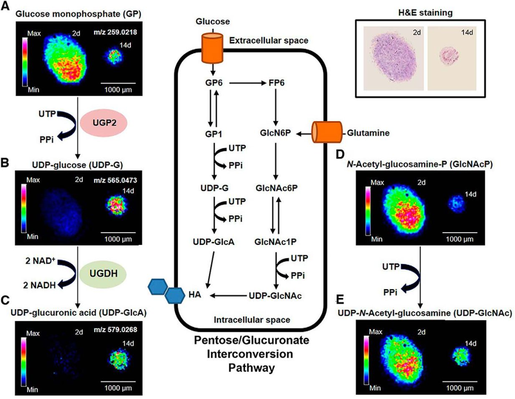
One of the applications that is gaining more and more attention is the use of MSI in the field of regenerative medicine. For example, in osteoarthritis (OA) pathology the impairment of cartilage regeneration can be related to a defective chondrogenic differentiation of mesenchymal stromal cells (MSCs). In a collaborative work with the INIBIC institute in Spain, we employed MALDI-MSI at several differentiation stages of human bone MSC spheroids. Our results revealed that UDP-glucuronic acid synthesis and amino sugar metabolism were downregulated in OA human bone MSCs during chondrogenesis compared to healthy cells. This work provided new potential therapeutic targets for OA.
https://pubmed.ncbi.nlm.nih.gov/31980557
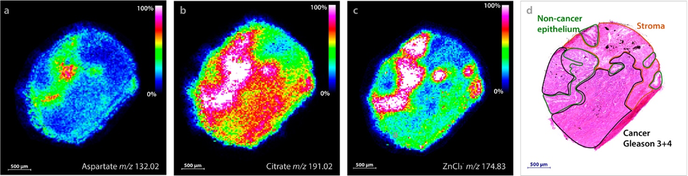
Another project in collaboration with the Tromsø University, allowed us to corelate the levels of zinc, citrate, and aspartate with a significant reduction in prostate cancer compared to non-cancer epithelium. The detection of these three molecules simultaneously, might provide a fast method for prostate cancer diagnostics and prognostics.
https://pubmed.ncbi.nlm.nih.gov/31944670
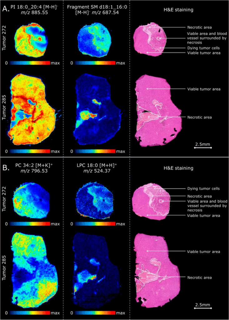
Another application in the field of oncology: Diffuse large B-cell lymphoma (DLBCL) is the most common B-cell non-Hodgkin lymphoma, and up to one-third of tumors ultimately relapse after treatment. In a collaboration with the BRIC institute in Copenhagen, we used a combination of in vivo DLBCL xenograft models and MSI to investigate intratumor heterogeneity. We identified specific lipid markers of viable and necrotic areas. Furthermore, we could monitor metabolic changes and found reduced adenosine triphosphate and increased adenosine monophosphate in tumors that were resistant to the chemotherapy treatment.
https://pubmed.ncbi.nlm.nih.gov/30422637
MSI is capable of simultaneously detecting thousands of different molecules in a spatially-resolved manner. In this way, the information obtained is more precise compared to data obtained by conventional mass spectrometry where typically, tissue extracts are used. In this way, the combination of MSI with other imaging modalities such as immunohistochemistry allows the incorporation of pathology information which is key in clinical studies.
Laser capture microdissection, proteomics, lipidomics, histology and data co-registration capabilities.
The M4i is a world leader in high resolution molecular imaging of biological surfaces, with concerted research efforts on three topics:
For this unique combination we host many guest researchers every year from different disciplines as part of the CORE lab. Our aim is to offer an open and collaborative environment where research and education meet.
How to apply:
All scientists, regardless of their affiliation, area of expertise or field of activity can benefit from Euro-BioImaging’s pan-European open access services. Potential users of these new technologies are encouraged to submit project proposals via our website. To do so, you can LogIn to access our application platform, choose the technology you want to use and the facility you wish to visit, then submit your proposal. All applications will be processed by the Euro-BioImaging Hub. As usual, users will benefit from advice and guidance by technical experts working at the Nodes, training opportunities, and data management services.
For more information: info@eurobioimaging.eu
July 9, 2026
In April 2026, the Med-Hub Head of Operations Alessandra Viale attended the 2026 180°N Conference, held at the beautiful Nye Hjorten Teater…
July 7, 2026
Armed conflicts generate long-lasting environmental contamination that extends well beyond the duration of military operations. The release of heavy metals such as Arsenic,…
July 6, 2026
On 2 July 2026, Euro-BioImaging hosted the online EVOLVE workshop “Building High-Quality Preclinical Imaging Facilities”, bringing together approximately 40 imaging facility staff and…