At the Special Edition Virtual Pub, "Imaging Micro- and Nanoplastics," Euro-BioImaging users, Node experts and Industry partners will explore how advanced microscopy, clinical imaging, and image analysis can be used to detect and study micro- and nanoplastic particles.
At this event, Jonathan Brewer, DaMBIC, Danish BioImaging Node, will reveal how fingerprint-region hyperspectral SRS and STED microscopy provide complementary chemical and spatial information, enabling label-free polymer identification together with high-resolution localization of nanoplastics in biologically relevant samples.
Join us on Friday, November 13, from 1-3 pm CEST for the Special Edition Virtual Pub!
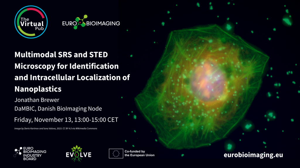
Multimodal SRS and STED Microscopy for Identification and Intracellular Localization of Nanoplastics
Speaker: Jonathan Brewer
Nanoplastics are increasingly implicated in human and environmental exposure scenarios, yet polymer-resolved detection inside cells remains challenging because extraction-based analytics lose spatial context and many plastics lack intrinsic fluorescence. We present a multimodal imaging workflow combining hyperspectral stimulated Raman scattering (SRS) microscopy with fluorescence and stimulated emission depletion (STED) microscopy for chemical identification and subcellular localization of nanoplastics in a mammalian cell model. Fingerprint-region SRS was performed on a BondXplorer platform using a fixed 1025 nm Stokes beam and pump scanning to cover ~661–1872 cm⁻¹. In mixed polymer samples, single-band SRS images and ROI-extracted spectra discriminated polystyrene (PS), polyethylene (PE), and polymethyl methacrylate (PMMA). Differentiated Caco-2 monolayers were then exposed to 500 nm PS beads. SRS imaging at the PS ring-breathing band (~1001 cm⁻¹) revealed punctate particle-associated signals, while ROI spectra recovered the characteristic PS fingerprint, providing in situ chemical confirmation. Correlative fluorescence and orthogonal SRS reslices supported localization of the particles within the cellular volume, while STED microscopy provided complementary super-resolution localization of individual nanoparticles within cells. Together, fingerprint-region hyperspectral SRS and STED microscopy provide complementary chemical and spatial information, enabling label-free polymer identification together with high-resolution localization of nanoplastics in biologically relevant samples.
More news from Euro-BioImaging
October 16, 2026
Interactive effects of microplastics and silver nanoparticles on onion roots: uptake, localisation and ROS detection via FLIM microscopy
At the Special Edition Virtual Pub, “Imaging Micro- and Nanoplastics,” Euro-BioImaging users, Node experts and Industry partners will explore how advanced microscopy, clinical…
From microplastics to pixels: open-source image analysis tools to study pollutant effects in zebrafish
At the Special Edition Virtual Pub, “Imaging Micro- and Nanoplastics,” Euro-BioImaging users, Node experts and Industry partners will explore how advanced microscopy, clinical…
Single-Shot Multiplex SRS Microscopy for Label-Free Imaging, Identification, and Quantification of Microplastics
At the Special Edition Virtual Pub, “Imaging Micro- and Nanoplastics,” Euro-BioImaging users, Node experts and Industry partners will explore how advanced microscopy, clinical…