Palina Nepachalovich is a first year PhD student studying lipid metabolism at the Center of Membrane Biochemistry and Lipid Research at TU Dresden . She is part of Maria Fedorova’s Group of bioanalysts, who use high resolution mass spectrometry and bioinformatics to study how living cells adapt to various stress conditions with a focus on lipids and lipid metabolism. Palina had never used a microscope when she applied for access at Euro-BioImaging’s Sofia Node in December 2021, as part of the Euro-BioImaging pilot User Access fund. Her promising project proposal, involving “Membrane disruptions and repair in cell death processes,” was selected for funding. We spoke to her while she was in Sofia, to learn more about what she hopes to accomplish and why this visit to our Sofia Node is so important to her research – and her career.
“At my home institution, my group does not generally use microscopy techniques. Our key expertise is mass spectrometry-based profiling of lipids that are characteristic for particular tissues, cells or subcellular organelles. We can determine what happens to lipid collectives both qualitatively and quantitatively upon various stress conditions. For example, we can find lipid fingerprints of cell death such as ferroptosis, which is mediated via oxidative damage of lipid membranes. But our particular methods do not allow us to observe how lipid membranes are damaged and repaired in real time. Fluorescence microscopy is no doubt a superior technique for obtaining a mechanistic view of these dynamic processes,” says Palina. “As a part of my PhD project, I am interested in the machinery of plasma membrane repair following damage. Due to its high temporal resolution, spinning disc confocal microscopy has a clear advantage in capturing these very fast processes.”
“One of my colleagues in Dresden told me about Dr. Stoynov’s team in Sofia. We thought it would be really cool to adapt their approach for DNA repair to study plasma membrane repair. As a Euro-BioImaging Node, they provide open access to their spinning disc confocal microscopes, full user training, and on top of that, they have developed a specialized image analysis software called CellTool to calculate the kinetics of protein behavior, which I also use in my project.”
Preparing for the trip
When Palina learned her project had been selected for funding in February 2022, she immediately went about making arrangements for her trip. The most critical part was getting specific cell lines expressing membrane repair proteins tagged with a fluorescent marker, making it possible to track them in living cells. For her project, several cell lines with tagged proteins of interest were kindly provided by the Hyman lab at MPI CBG in Dresden.
Microscopy crash course
She finally arrived in Sofia in July 2022. From Day 1, she was able to sit at the microscope. “In just a couple of days, the Sofia team trained me. I was really impressed by how they could explain in really easy words what is going on with the instrument and how to set it up. I especially want to thank Radoslav Aleksandrov, Peter Kanev and Ivana Roeva for their support and help with my experiments. Now I am using the instruments without any assistance, but of course I can ask for advice any time I need it,” she says proudly.
After an initial fast progression, the second two weeks of her stay were spent on optimization. “The introduction to practical microscopy is very general, but what I’m doing is very specific, with a number of fluorescent probes each tagging a particular cellular events. It was really crucial for me to dig into different live cell imaging protocols and procedures,” she explains.
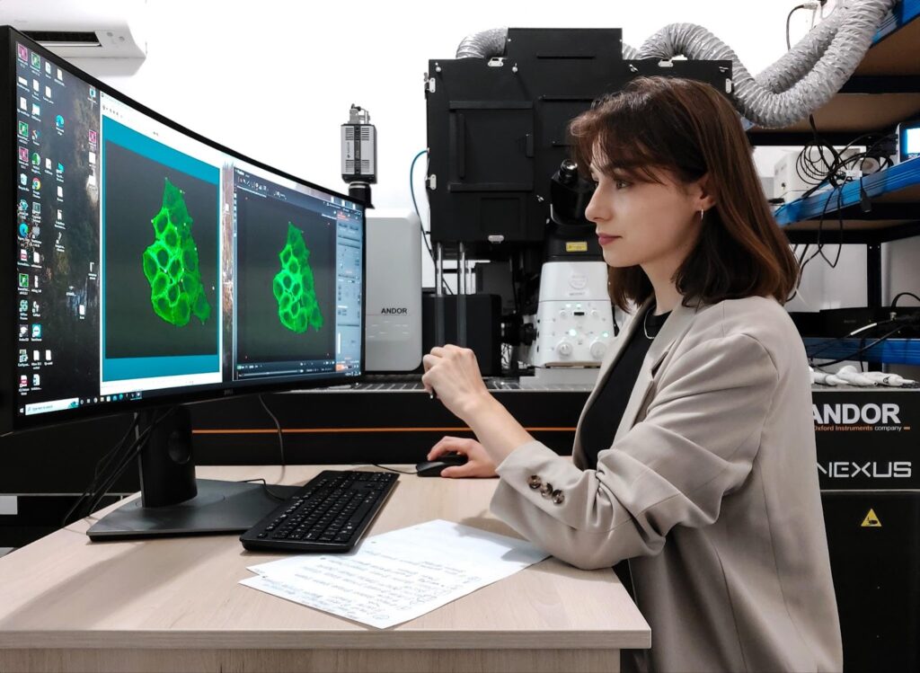
Palina Nepachalovich acquires data using spinning disc microscopy at Euro-BioImaging’s Sofia Node in Bulgaria.
Sharing expertise
“The team in Sofia provides invaluable technical advice. We are adapting their research from DNA repair to plasma membrane repair. I follow their approach, using laser-triggered plasma membrane rupture almost in the same way they trigger the damage of DNA. So I really benefit from their expertise. They give me a lot of advice on how to adapt the laser, how to adjust the camera, etc..”
Challenges of working on membrane repair
It is quite challenging to work on membrane repair. The plasma membrane is very thin and the damaged membrane seals in seconds. Palina has to be concentrated and move quickly. “It is within a very short timespan that the membrane repair machinery acts at the site of the damage and the repair happens,” she explains. “That’s why I really need the high temporal resolution,” she explains.
She has finally found the optimal settings for live cell imaging of this super fast process. She now needs to screen the cell lines expressing different candidate proteins to understand which role they play in the membrane repair process. The timing is tight. “Even when everything is set up, it still takes a full day to get 50 time lapse movies of the repair process in every single cell line.”
“This experience has been invaluable. In Sofia, not only did I receive training on how to use the machine with the focus on my custom project, but the team supports me constantly with their really specific technical expertise, and I have nearly unlimited time for my experiments.”
Expected outcomes
Although her time in Sofia is nearly over, this is just the beginning of Palina’s exploration. When she returns to Dresden, she will test her approach on many more cell lines. “I will return with a large knowledge base about what I should do. After this research stay, I can go directly to a microscope facility and continue my research on my own with the things I learned from the team in Sofia.”
Scientifically, the outcome of Palina’s experiments are yet to be seen and analyzed in depth. But a very important outcome has already been achieved. Palina has discovered the power of spinning disc microscopy as applied to her field of research. And this is right in line with Euro-BioImaging’s mission – to make state of the art imaging approaches available to all scientists, no matter their area of expertise.
About the Sofia Node
The Sofia BioImaging Node is a multimodal single-sited Node, located at the Institute of Molecular Biology, Bulgarian Academy of Science, in Sofia. The Bulgarian Center for Advanced Microscopy at IMB-BAS is included in the National Roadmap for Scientific Infrastructure and as part of Euro-BioImaging provides open user access to a range of state-of-the-art technologies in biological and biomedical imaging for life scientists. With a specific focus on fast and high-resolution live-cell imaging and laser-based disruptions of cells and their components, the Node provides outstanding expertise in the application of Spinning Disk microscope systems and the use of UV laser micro-irradiation and FRAP to study protein dynamics and DNA repair. For more information see the Bulgarian Node Home page..
More news from Euro-BioImaging
July 20, 2026
Strengthening research-industry partnerships: results from the EVOLVE industry job shadowing pilot
The EVOLVE project, supported by European Union funding, recently completed a pilot programme designed to deepen the relationships between academic research infrastructures…
Imaging-Driven Neuroscience, Responsible Research, and the Power of Advocacy: Highlights from FENS Forum 2026
Euro-BioImaging was pleased to participate in the FENS Forum 2026, Europe’s largest international gathering dedicated to advancing neuroscience (>8000 participants). Throughout…
Euro-BioImaging Highlights the Value of Research Infrastructures at the FENS 2026 Satellite Symposium hosted by EBRAINS
Euro-BioImaging was pleased to participate #FENS2026 Satellite Symposium “Accelerating Your Neuroscience Research through the European Research Infrastructures” organised by EBRAINS, to learn…