
April 28, 2025
Beyond the husk – shining light on seed quality through molecular imaging
Seed quality is absolutely key to the success of both agronomically relevant crops and forest tree species used for restoration strategies. Forest restoration…
April 28, 2025
Seed quality is absolutely key to the success of both agronomically relevant crops and forest tree species used for restoration strategies. Forest restoration…
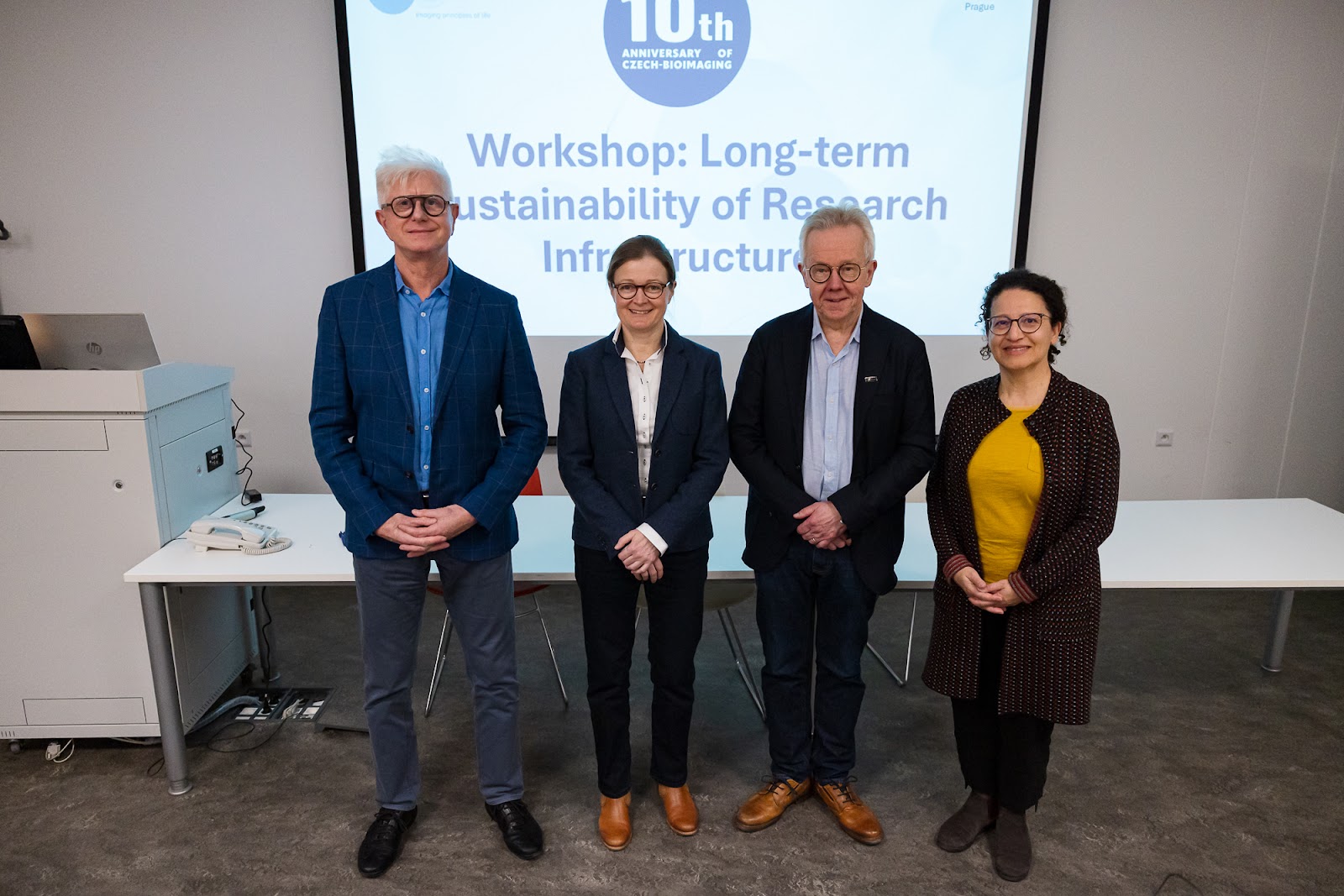
April 25, 2025
In 2025, Czech-BioImaging celebrates its 10th anniversary. Czech-BioImaging is the national infrastructure that brings together 16 leading imaging centers across the Czech…

April 24, 2025
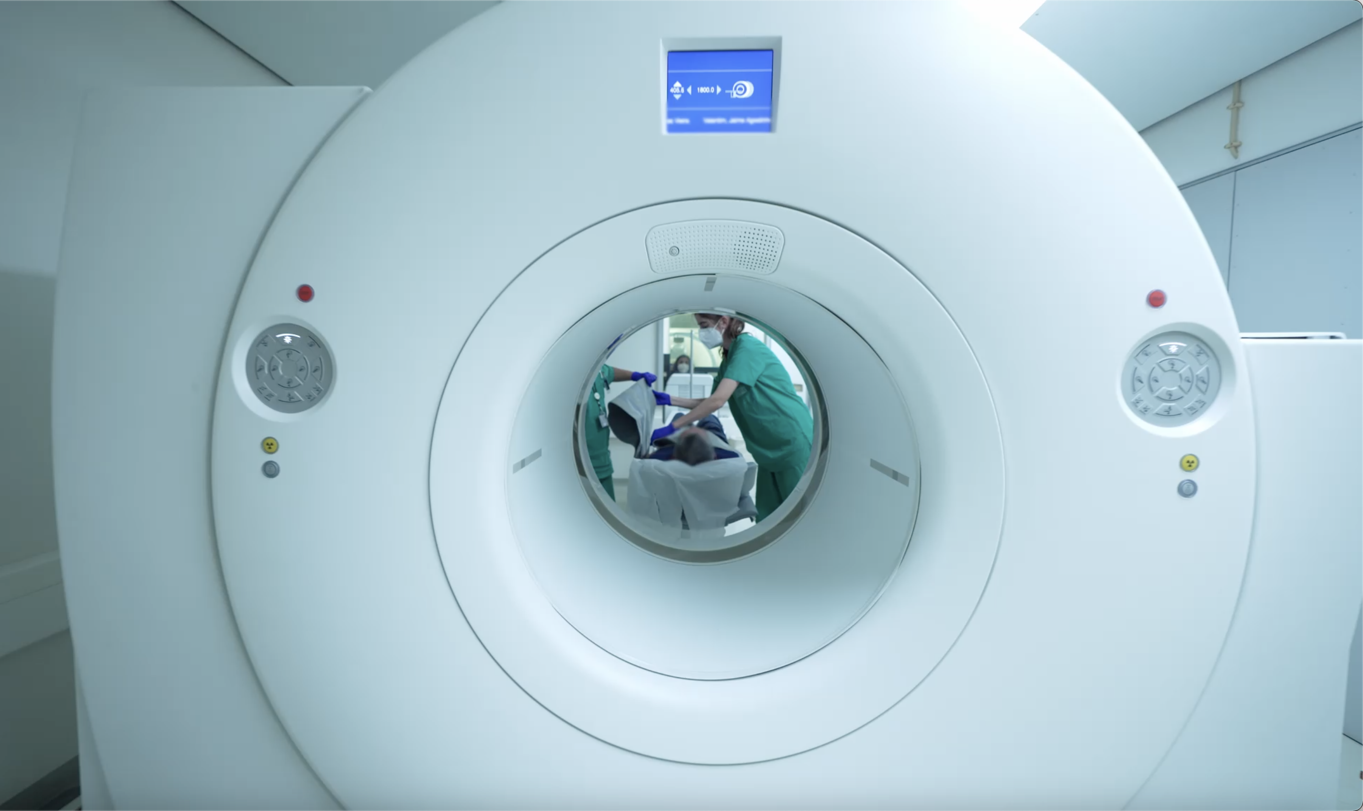
Colorectal cancer deaths are rising among young adults. Rabe’ah Almahassneh, a PhD student from the University of Valencia who is developing her research…

April 17, 2025
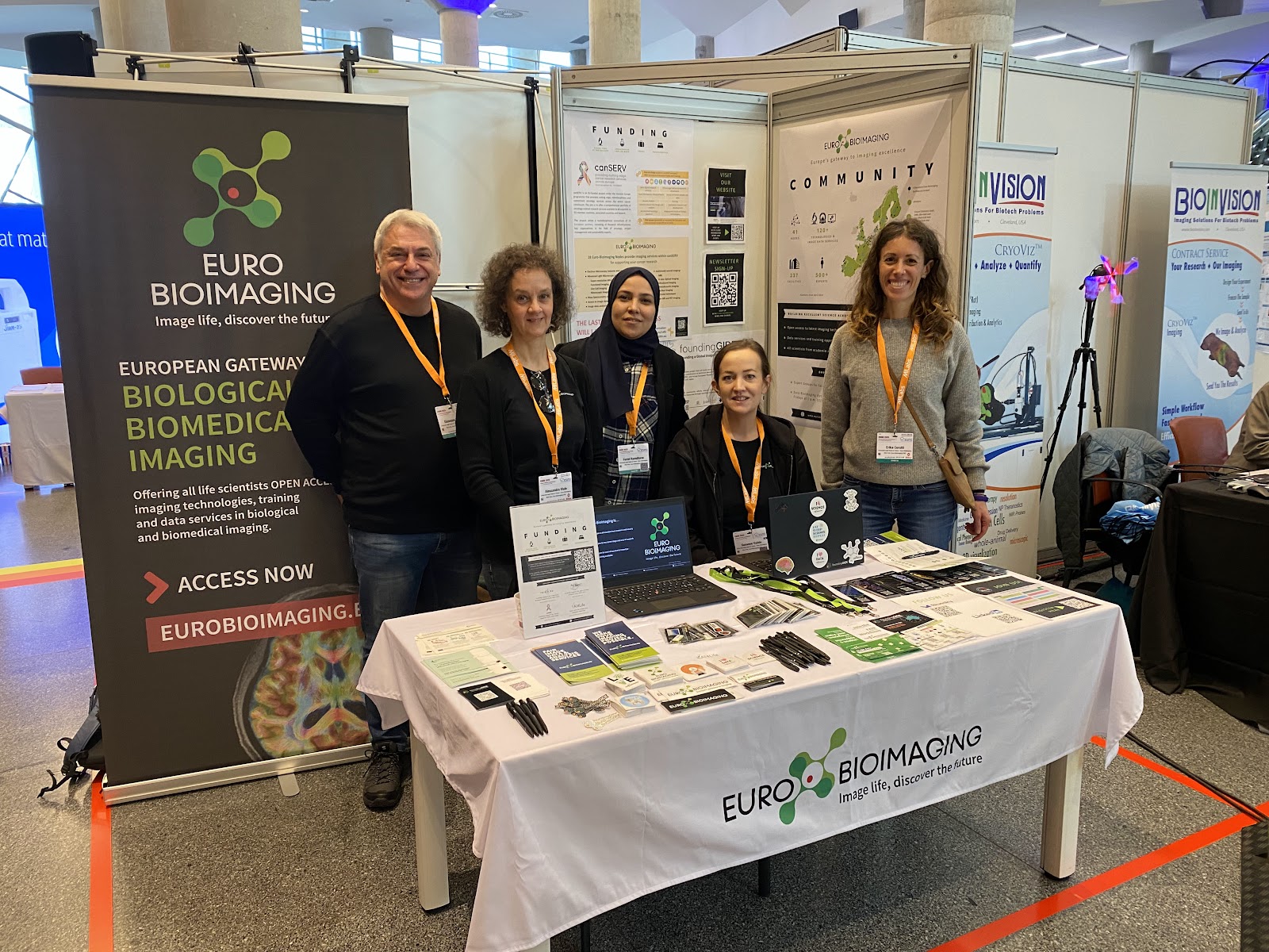
The Euro-BioImaging Bio-Hub team represented Euro-BioImaging with a booth at the German Biotechnology Days 2025, which took place on 9th and 10th April…
April 17, 2025
In a compelling EVOLVE Mentoring Masterclass hosted by Euro-BioImaging, Professor Ilaria Testa offered a multifaceted look into her scientific journey, from her interdisciplinary…
April 16, 2025
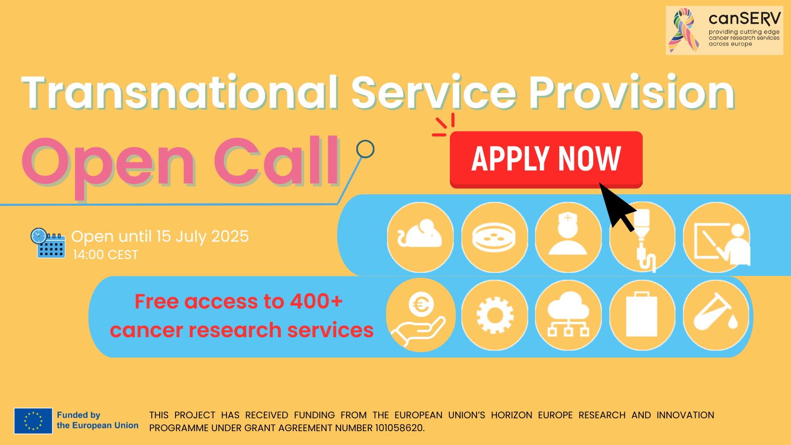
Euro-BioImaging is pleased to announce that the fourth and final Open Call from the Horizon Europe-funded canSERV project is here! Cancer Researchers are…

April 9, 2025
From March 25-28, Euro-BioImaging Hub team was delighted to host our Node community & industry partners. The Euro-BioImaging All Hands Meeting is the…
April 9, 2025
EIC Summit 2025 Last week, Strategic Business Developer Claudia Pfander and Anne-Charlotte Joubert, Senior Officer for European Affairs, represented Euro-BioImaging at the…
April 9, 2025
Euro-BioImaging staff was delighted to attend the European Molecular Imaging Meeting 2025 in Bilbao that took place from March 11th to March…

April 8, 2025
In early 2025, Euro-BioImaging Finland, the Finnish national infrastructure that brings together the Finnish Advanced Microscopy Node (FiAM) and the Finnish Biomedical Imaging…
April 8, 2025
Clinical evaluation of novel treatments and comparisons with existing ones is of paramount importance in the development of drugs and therapies, to ensure…

April 8, 2025
Participate in the Four Seasons of the Invisible, Euro-BioImaging’s imaging contest! Join us to celebrate the four seasons seen through the prism of…