
April 7, 2025
Towards the development of novel SARS-CoV-2 therapeutics
Dr. Veronika Huntosova and her team at the Center for Interdisciplinary Biosciences, Technology and Innovation Park, Pavol Jozef Šafárik University in Košice, Slovakia,…
April 7, 2025
Dr. Veronika Huntosova and her team at the Center for Interdisciplinary Biosciences, Technology and Innovation Park, Pavol Jozef Šafárik University in Košice, Slovakia,…

April 4, 2025
Do you have an innovative research project in the field of biomedical imaging? Czech-BioImaging offers funding opportunities to support the best research ideas,…

April 4, 2025
Linda Krahula Doleží is an assistant professor at the Language Centre at the Masaryk University in Brno. She did her PhD on Czech…

April 4, 2025
In early 2024 the first cohort of Euro-BioImaging Scientific Ambassadors was onboarded. Twelve highly enthusiastic researchers were chosen to represent Euro-BioImaging and raise…

March 28, 2025
We are excited to announce that the paper “Enabling Global Image Data Sharing in the Life Sciences” has been published in Nature Methods…

March 27, 2025
Euro-BioImaging, alongside Simula, EPFL and the University of Bergen, is proud to announce the launch of FAIR Image Analysis Across…

March 27, 2025
We are delighted to announce that the MultiModal Molecular Imaging Italian Node and the Advanced Light Microscopy (ALM) Italian Node are launching a…

March 24, 2025
Between 20-40% of global food crop production is lost each year due to plant pests and pathogens, highlighting the urgency for scientific research…
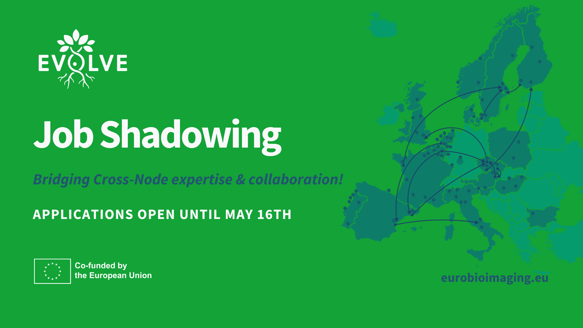
March 24, 2025
Are you a Euro-BioImaging Node staff member eager to broaden your professional horizons, exchange innovative ideas, and enhance your expertise? The second call…
March 20, 2025
The European Commission (EC) Directorate-General for Research and Innovation (DG RTD) has recognised the AI4Life project, coordinated by Euro-BioImaging, for its achievements, selecting…
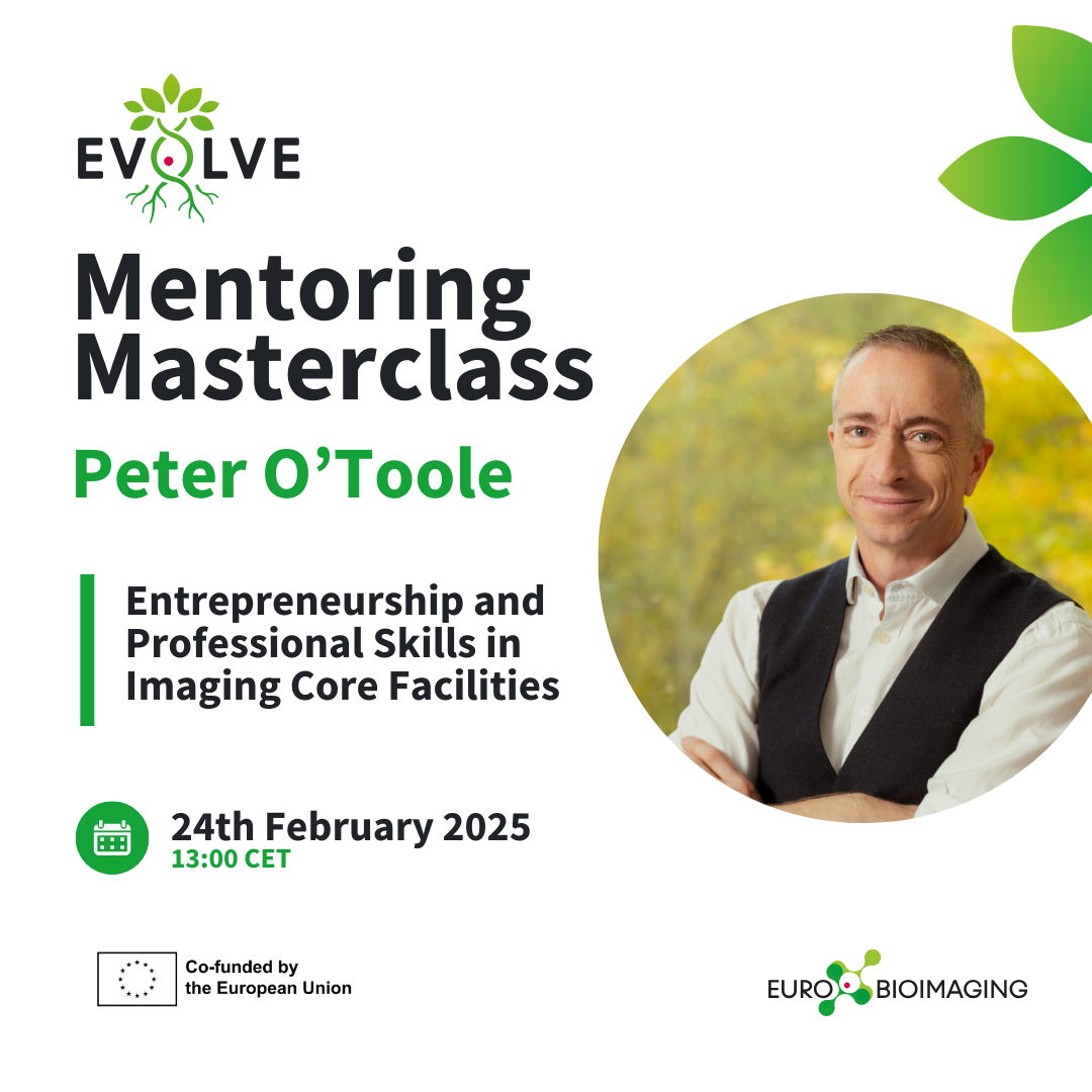
March 14, 2025
The EVOLVE Team2025 Mentoring Masterclass series kicked off with an engaging session featuring Peter O’Toole, President of the Royal Microscopical Society and Director…

March 13, 2025
At this year’s BioHackathon Europe 2024 in Barcelona, a dedicated group of enthusiasts developed FAIR image analysis workflows in the Galaxy platform. Led…