October 30, 2023
Euro-BioImaging opens a Call for Node Upgrades
Euro-BioImaging ERIC is pleased to announce that its Call for Node Upgrades 2023 opens on November 1! This is the process by which new…
October 30, 2023
Euro-BioImaging ERIC is pleased to announce that its Call for Node Upgrades 2023 opens on November 1! This is the process by which new…
October 25, 2023
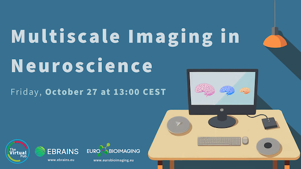
Our next Special Edition Virtual Pub, “Multiscale Imaging in the Neurosciences,” organized in collaboration with EBRAINS, will take place on Friday, October 27. At…
October 25, 2023
Our next Special Edition Virtual Pub, “Multiscale Imaging in the Neurosciences,” organized in collaboration with EBRAINS, will take place on Friday, October 27. At…
October 25, 2023
Our next Special Edition Virtual Pub, “Multiscale Imaging in the Neurosciences,” organized in collaboration with EBRAINS, will take place on Friday, October 27. At…
October 25, 2023
Our next Special Edition Virtual Pub, “Multiscale Imaging in the Neurosciences,” organized in collaboration with EBRAINS, will take place on Friday, October 27. At…
October 25, 2023
Our next Special Edition Virtual Pub, “Multiscale Imaging in the Neurosciences,” organized in collaboration with EBRAINS, will take place on Friday, October 27. At…

October 16, 2023
We are delighted to announce that Euro-BioImaging has launched a Scientific Ambassador program. This program is designed to raise awareness about the services and…
October 13, 2023
We are delighted to share the program for our Special Edition Virtual Pub “Multiscale Imaging in the Neurosciences,” organized in collaboration with EBRAINS. This…
October 12, 2023
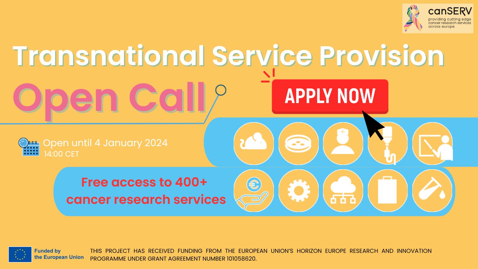
Euro-BioImaging is pleased to announce that the first Open Call from the Horizon Europe-funded canSERV project is here! Cancer Researchers are invited to apply…
October 6, 2023
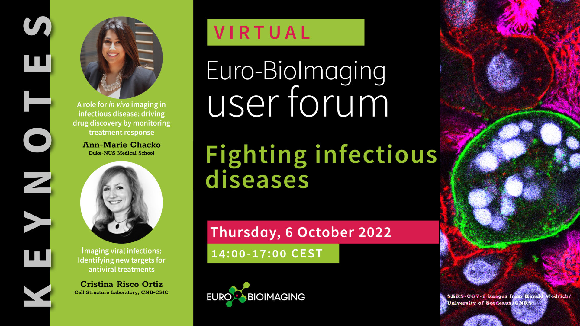
Euro-BioImaging is organizing a fourth online User Forum on Thursday, October 6, 2022 from 14:00-17:00 CEST. The topic is “Fighting Infectious Diseases.” This event…
October 2, 2023
The EUCAIM consortium and the European Commission are excited to announce the first public release of its platform, Cancer Image…

September 22, 2023
Within the framework of the COMULISglobe project, we will be awarding a total of 4 grants for access to Euro-BioImaging…