July 1, 2026
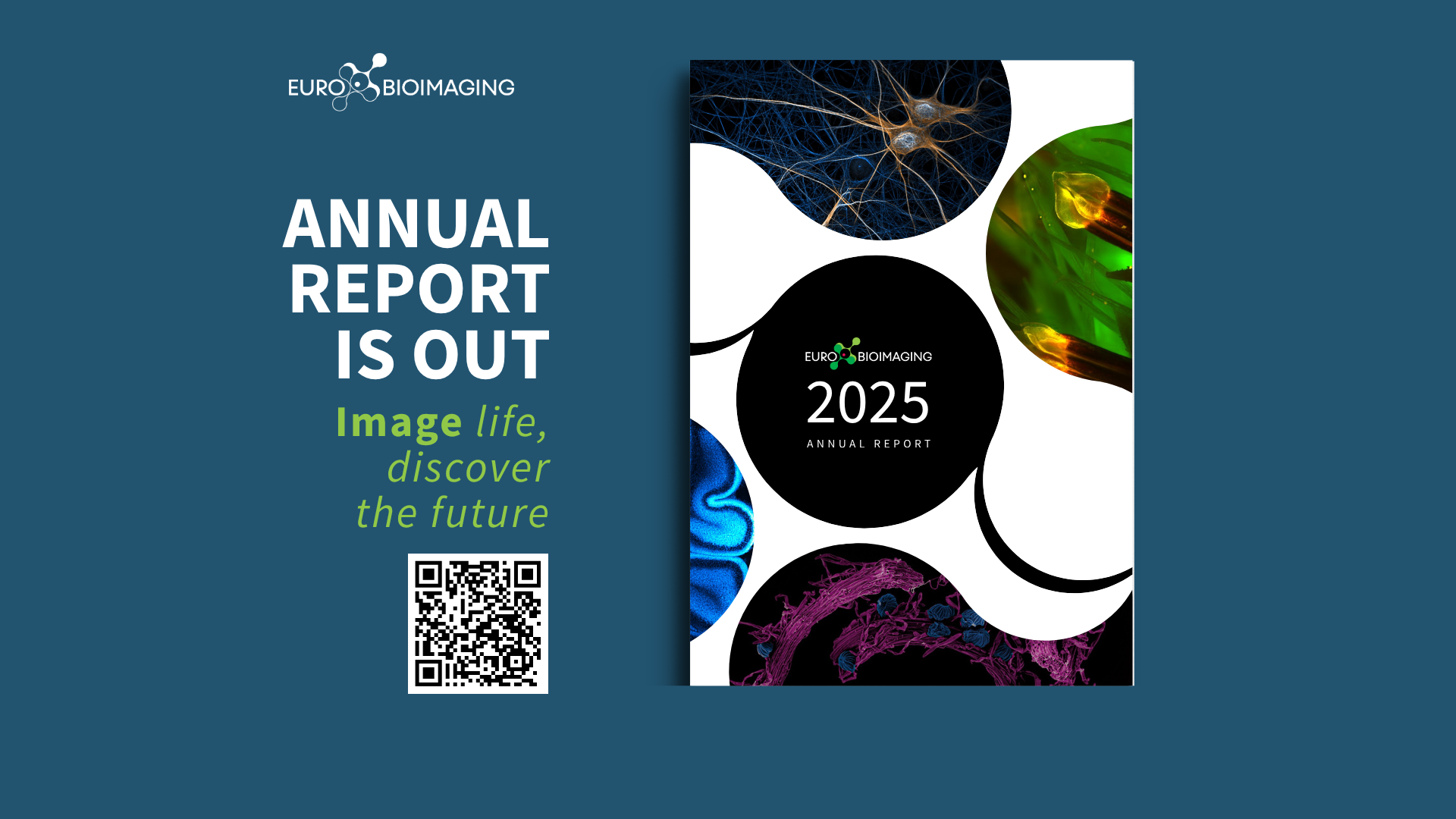
Introducing the Euro-BioImaging Annual Report 2025
The Euro-BioImaging Annual Report 2025 is now online! It shares our highlights from 2025, from expanding our community to strengthening our impact, and…

Euro-BioImaging is organizing an online User Forum on April 5, 2022, from 14:00-17:00 CEST. This event will highlight the importance of cutting-edge imaging technologies in support of brain research and showcase the specific expertise available at our Nodes across Europe through case studies presented in tandem with the research community.
In this abstract, learn how Angela Getz and her colleagues at the Bordeaux Imaging Center (part of the French BioImaging Node) and the Interdisciplinary Institute for Neuroscience in Bordeaux, combined advanced imaging tools, such as lattice light sheet and photomanipulation, with new molecular tools for visualizing and controlling the dynamics of AMPA receptors to understand synaptic plasticity.
Hear this talk and others like it on April 5 at the Euro-BioImaging User Forum: At the Forefront of Neuroscience.
Studying AMPA Receptor Dynamics with Lattice Light Sheet and Multiphoton Microscopy
Angela Getz, Bordeaux Imaging Center/IINS
Daniel Choquet, France BioImaging
The focus of this study is the contribution of AMPAR membrane diffusion/trapping to short- and long-term synaptic plasticity, investigated using a combined developments in advanced imaging with new molecular tools for visualizing and controlling AMPAR surface diffusion to understand the respective roles of presynaptic neurotransmitter release scaling and postsynaptic AMPAR dynamics in tuning synaptic plasticity.
To image endogenous, fluorescently labelled AMPA receptors in live organotypic hippocampal brain slices, a home-made lattice light-sheet (LLS) microscope was used. LLS allows fast and non-phototoxic imaging with subcellular resolution in live 3D samples. The system was modified with the addition of a targeted photo-stimulation module to optically manipulate the sample with high spatiotemporal resolution, where visible and fs NIR lasers were coupled to the photo-manipulation path. One-photon targeted photobleaching was used to measure endogenous AMPAR surface diffusion by FRAP. Using two-photon photostimulation, synaptic Ca2+ transients were observed using glutamate uncaging with the genetically encoded calcium sensor GCaMP6f, and 4D structural plasticity was monitored via spine volume changes using photoactivatable CaMKII. To probe pre-synaptic transmitter release, a multimodal two-photon microscope was used, which is equipped with 2 scanners allowing to couple imaging and photostimulation, 3 fs NIR lasers, and synchronized with an electrophysiology rig. Pre-synaptic glutamate release, triggered by electrical stimulation, was observed on live organotypic and acute mouse brain slices using the fluorescent glutamate sensor iGluSnFR and a fast linescan imaging protocol (500Hz) to temporally resolve how fast neurotransmitter release events change during activity-dependent short-term plasticity.
These developments are allowing the study of the molecular mechanisms underlying the spatiotemporal expression of synaptic plasticity in integrated networks. This work demonstrates the high level of instrumentation and staff skills present at the BIC targeted to neuroscience research and open to the whole community.

July 1, 2026
The Euro-BioImaging Annual Report 2025 is now online! It shares our highlights from 2025, from expanding our community to strengthening our impact, and…

June 26, 2026
Euro-BioImaging, represented by Antje Keppler, Director of the Euro-BioImaging ERIC Bio-Hub, was pleased to contribute to the Irish Universities Association’s Future of Ireland event in…
June 26, 2026
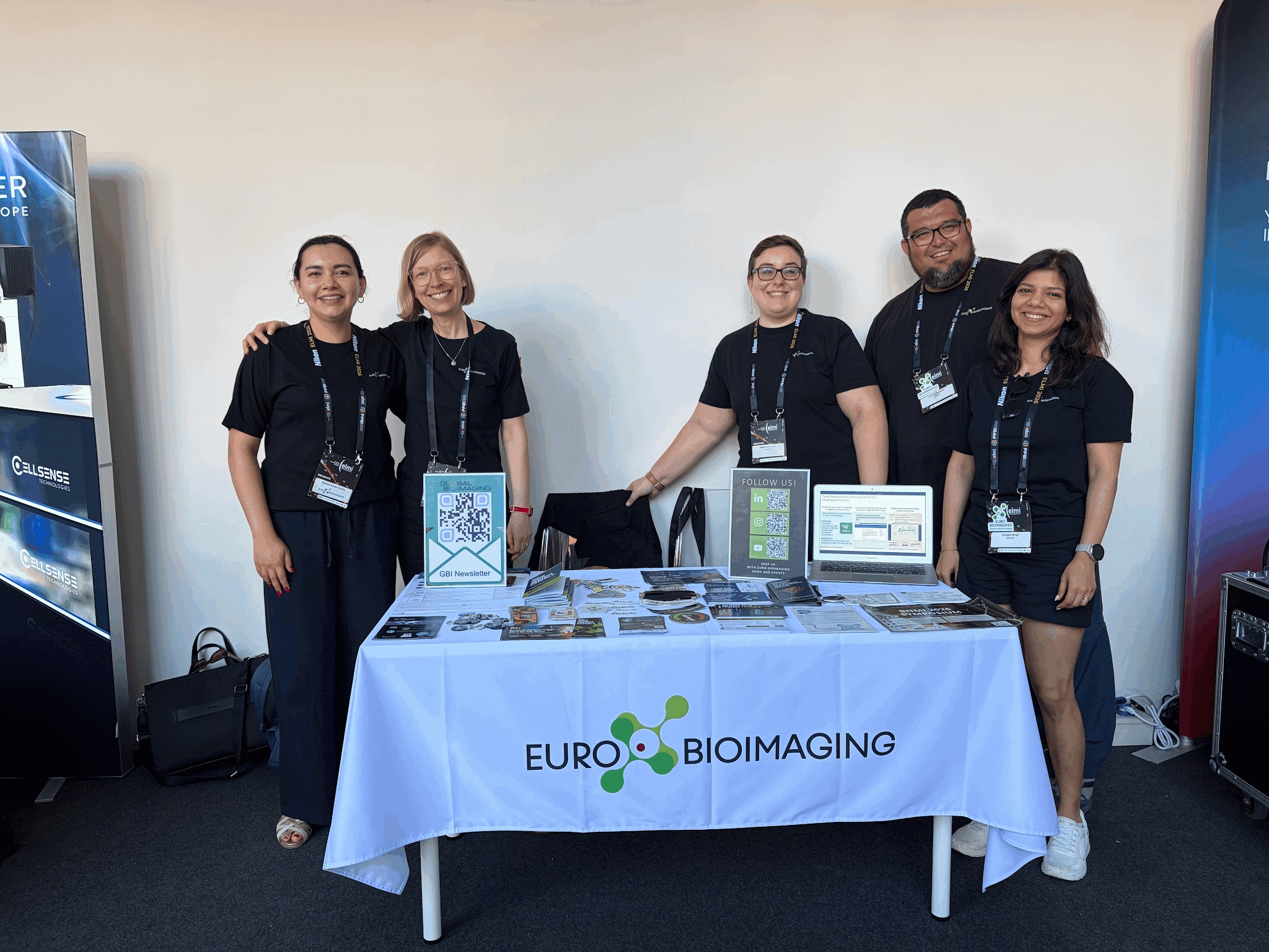
The annual meeting of the European Light Microscopy Initiative (ELMI) took place in Coimbra, Portugal, from June 16-19th, 2026. With nearly 600 in…