May 23, 2025
Imaging for understanding plant disease dynamics
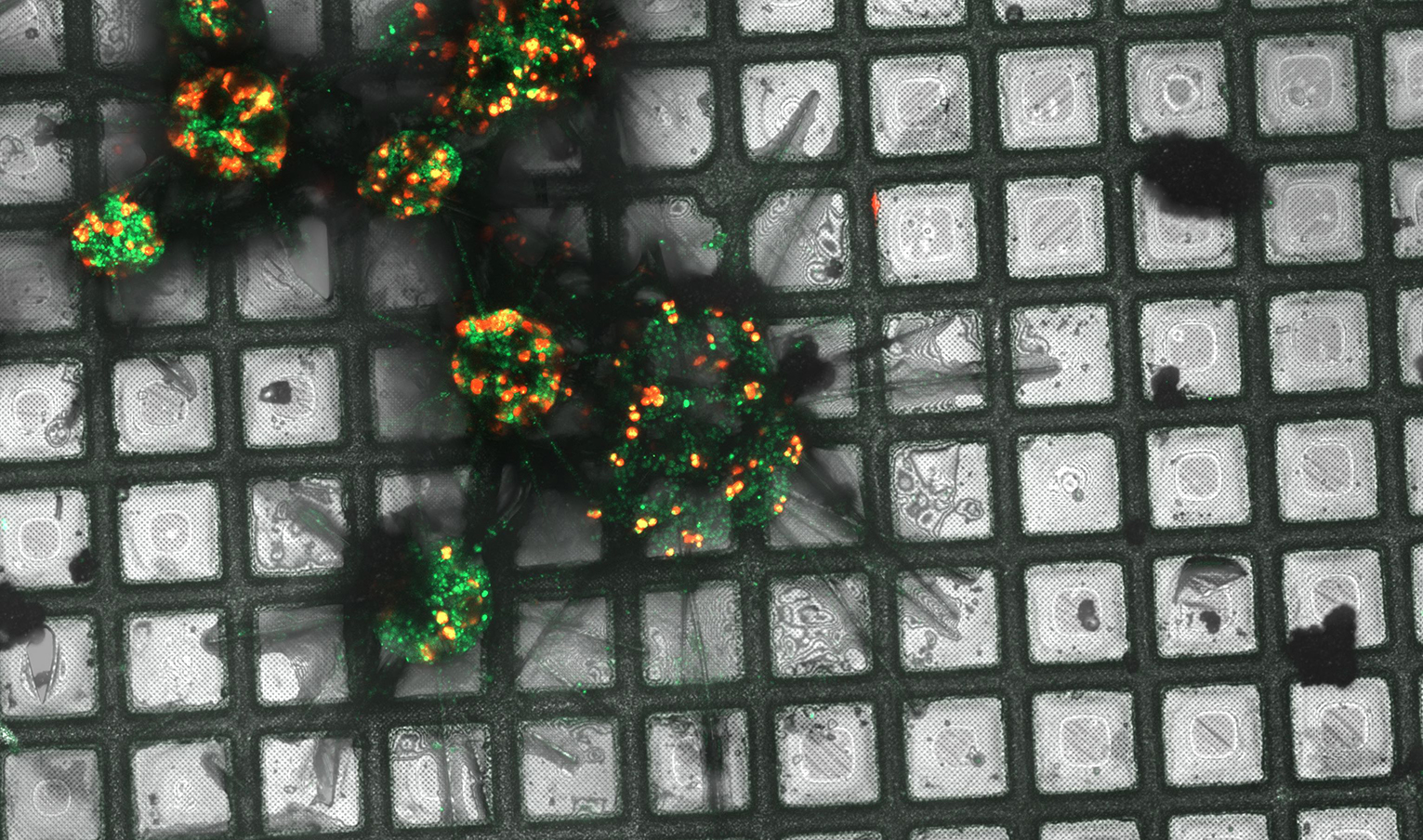
Xylella fastidiosa is a vector-borne gram-negative xylem-limited bacterium native to the Americas, affecting more than 650 plant species, including crops of major economic importance. The bacterial sequence…