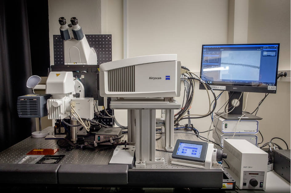
Imaging living plants has always been challenging. Most microscopes place the sample horizontally – while plants grow vertically. But scientists at Czech Republic’s Institute of Experimental Botany, part of Euro-BioImaging’s Advanced Light and Electron Microscopy Node Prague, have found a way to overcome this challenge. Inspired by scientist in IST Vienna, they’ve turned their Zeiss LSM 880 with Airyscan detector on its side – at an angle of 90 degrees – and added high magnification immersion objectives - for fast and sensitive imaging of root growth in its natural (gravitational) orientation. With long-term experience in plant biology, and the right combination of high-end instruments and expertise, our Node in Prague is optimized for live plant studies. And the technologies and expertise they offer areavailable to all scientists via the Euro-BioImaging portal. Learn more in this interview with Kateřina Malínská, head of the Imaging Facility at the Institute of Experimental Botany.
Your institute has “toppled” a confocal laser scanning microscope to specifically observe plant development. Please tell us a bit more about the capabilities of this system.
The root tip inspected by classical microscopes attempts to bend downwards, which is not possible in this setup. The plant is therefore permanently gravi-stimulated during all measurements. This might influence the data obtained by possible unwanted artifacts and makes some experiments on growing roots completely impossible. In 2019, inspired by IST Vienna (see article on Euro-BioImaging website here), local representatives of the Zeiss company have toppled Zeiss LSM880 with Airyscan detector to optimize it for our plant research. In this setup, vertical sample mounting preserves the natural direction of plant growth and enables long-term experiments on plants acquired with confocal resolution. In addition, the gravity vector can be finely tuned by a rotational stage insert, which allows the application of the desired gravi-stimulus. Compared to the previously established setup in Vienna, we have used Airyscan super-resolution detector and installed high magnification immersion objectives (63x and 100x). This allowed imaging in much higher spatial resolution. The long-term imaging opened new challenges that had to be optimized subsequently. The root growth had to be compensated by microscope stage movement and the plant leaves illuminated to preserve their photosynthetic activity. These two aspects were fixed during the last year and are still being improved.
Are there any particular challenges with this new microscope orientation? What things do you have to pay special attention to?
During the installation, there were several technical issues that had to be solved. The microscope body was rotated and fixed to the anti-vibration table by a holder. The scanning head could preserve its original orientation due to the elevated mounting but its side cover had to be replaced by the 3D-printed cover not interfering with motorized stage movement. Since the elevated eyepieces were not easily accessible, an additional AIO computer providing real-time eyepieces view taken by CCD camera was installed to increase user comfort. Two types of stage inserts were installed, the commercial one and the customized rotational insert that was gradually developed according to our needs.
A long-term vertical-stage imaging opened new challenges that had to be addressed, among them how to compensate for the root growth out of the field of view. After initial tile scanning, we had to adopt a more sophisticated solution. Now the root growth is compensated by microscope stage movement, allowing time-lapse imaging in a single frame. For this, Ph.D. student Ivan Kashkan, a member of our facility team, implemented Zen macro Microscopy Pipeline Constructor (MyPiC). MyPiC was developed in the group of Jan Ellenberg, EMBL, Heidelberg by Antonio Politi and Ivan adapted it for our LSM880 setup. It uses center of mass of fluorescence signal for the tracking and allows multi-position and multi-channel z-stack time-lapse experiments. The biggest challenge however, was to time-lapse root growth with high magnification or/and with Airyscan detector. Here we highly appreciate Matlab based TipTracker algorithm and close collaboration with Dr. Hauschild from Vienna IST Imaging & Optics Facility.
What kind of projects do you support using this technology? What scientific questions are you be able to address?
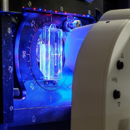
Fig. 2: Plant growing in coverglass chamber is illuminated during long-term experiment (blue-red light in the picture) and the fluorescence signal is acquired.
You also provide a range of expertise in other microscopy techniques. What other services do you provide in your facility and how are they useful in combination with this type of microscopy?
Our main focus is in vivo imaging of plants and our users can extend their research originally made on “classical” LSM880 (or other) system by imaging on vertical-stage LSM880. Similarities between both systems help users to perform experiments easily. To run long-term experiments, careful pre-selection of suitable seedlings is crucial. For this purpose, we placed the fluorescence stereomicroscope Leica next to the vertical-stage LSM880.
You can imagine a typical workflow at the vertical-stage microscope in several steps. The story usually starts with a long-term imaging with a dry low magnification objective. This is the crucial phase where the new phenomena are usually noted and their time and space windows in growing seedling are estimated. Then you can zoom in with higher magnification objectives (40x or 63x) and utilize the Airyscan detector providing high spatial resolution. Consequently, the observed effect can also be studied with our other microscopy systems, like spinning disk microscopy with ultrafast simultaneous dual channel imaging for studying highly dynamic processes or recently installed LSM900 with Airyscan2, which can provide both high speed and high resolution imaging in multiplexed mode. Moreover, we have equipped this system with sensitive EMCCD camera to allow parallel imaging of bioluminescent plants.
Why should scientists choose your facility to study plant biology?
With a long-term experience in plant biology, we are pleased to offer high-end instruments and expertise for challenging tasks in the field of non-invasive in vivo fluorescence microscopy with high spatial and temporal resolution. However, some of our users here also solve tasks from the medical or technical field. And of course, the vertical-stage LSM880 with Airyscan is ready for your experiments ;-).
About the Advanced Light and Electron Microscopy Node Prague
The Czech Advanced Light and Electron Microscopy Multi Modal Multi Sited Node located in Prague and in České Budějovice offers a wide range of state-of-the-art light and electron microscopy equipment and techniques, offered by five closely collaborating core facilities. The Node is also active in organizing training and courses focused on theoretical and practical aspects of basic and advanced microscopy techniques. Light microscopy instrumentation includes microscopy systems that range from multi-functional point and spinning disc confocal microscopes, light-sheet system up to high-end multi-photon and super-resolution microscopy systems. In the area of electron microscopy, the Node provides expertise and cutting-edge equipment for a broad range of biological sample preparation and ultrastructural imaging techniques, including some cryo techniques, electron tomography, SBF-SEM, FIB-SEM and CLEM. Broad expertise in experiment design and data acquisition is also complemented by extensive data analysis services. Learn more here.
Building Skills Across Europe: EVOLVE Supports the Second Edition of the Distributed Image Analysis Training Course
The second edition of the “Introduction to Image Analysis with Python for Life Scientists” course marked another successful milestone in the development of Euro-BioImaging’s distributed training model.
Strengthening research-industry partnerships: results from the EVOLVE industry job shadowing pilot
The EVOLVE project, supported by European Union funding, recently completed a pilot programme designed to deepen the relationships between academic research infrastructures…
Imaging-Driven Neuroscience, Responsible Research, and the Power of Advocacy: Highlights from FENS Forum 2026
Euro-BioImaging was pleased to participate in the FENS Forum 2026, Europe’s largest international gathering dedicated to advancing neuroscience (>8000 participants). Throughout…