
October 16, 2026
RAISE Hub: One gateway to Europe’s AI in Science Community
A new online platform to connect researchers, infrastructures, and funders to accelerate AI scientific discovery across Europe Dublin, Ireland, 15 October…



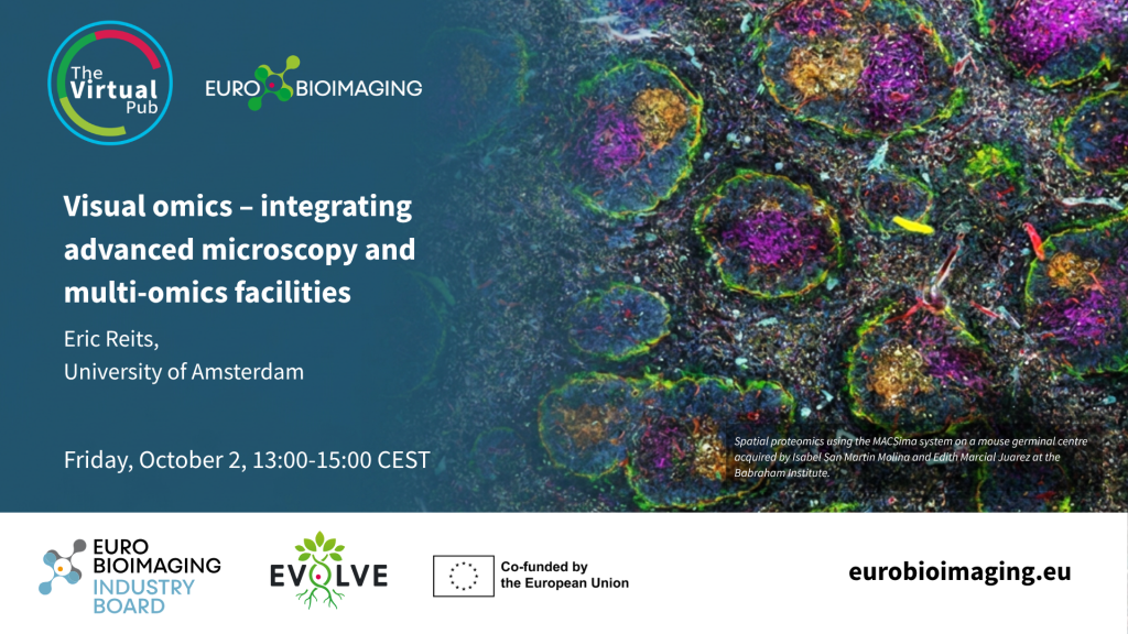
Spatial proteomics is rapidly transforming the way researchers study complex biological systems. To explore the latest developments in this fast-moving field, Euro-BioImaging is organising a Special Edition Virtual Pub dedicated to Spatial Proteomics on Friday, October 2, from 13:00–15:00 CEST, bringing together experts from academia and industry.
At this event, Eric Reits, University of Amsterdam, will tell us how the Dutch Large-Scale Research Infrastructures NL-BioImaging (including the Advanced NL-Microscopy Euro-BioImaging node) and the Netherlands X-omics Initiative (X-omics) are working towards combining advanced microscopy technologies and multi-omics with FAIR data management and analysis to serve users.
Visual omics – integrating advanced microscopy and multi-omics facilities
By: Eric Reits, University of Amsterdam
Visual omics is an exciting emerging research frontier, where the complex integration of advanced microscopy and omics facilities will lead to new integrated analysis methods. The Dutch Large-Scale Research Infrastructures NL-BioImaging(including the Advanced NL microscopy EuroBI node) and the Netherlands X-omics Initiative (X-omics) have built strong, complementary foundations with innovative technologies including advanced microscopy (e.g. high-resolution, timelapse and high-content microscopy) and multi-omics (e.g. genomics, proteomics and metabolomics), each supported and connected by a national FAIR data management and analysis node. Visual Omics is the next-generation integration of NL-BioImaging and X-omics, moving beyond static snapshots to "record" the molecular history of living cells. Ideally, the full range of microscopy techniques can be used with the full range of multi-omics techniques, linking the microscopy and omics core facilities within and between institutes, enabling integration of complementary imaging and omics technologies. However, both scientists and current infrastructure facilities face a number of challenges that have to be addressed at the national level in order to:
Enabling true integration of multi-scale imaging of cells, organoids, tissues, plants, and small animal models with multi-omics data will enable scientists to cross disciplines and push the boundaries of their chosen specializations, ranging from fundamental insights in biology to developing new strategies for societal challenges including agriculture and a variety of diseases including cancer, neurodegenerative, and cardiovascular diseases.
October 16, 2026
A new online platform to connect researchers, infrastructures, and funders to accelerate AI scientific discovery across Europe Dublin, Ireland, 15 October…
October 16, 2026
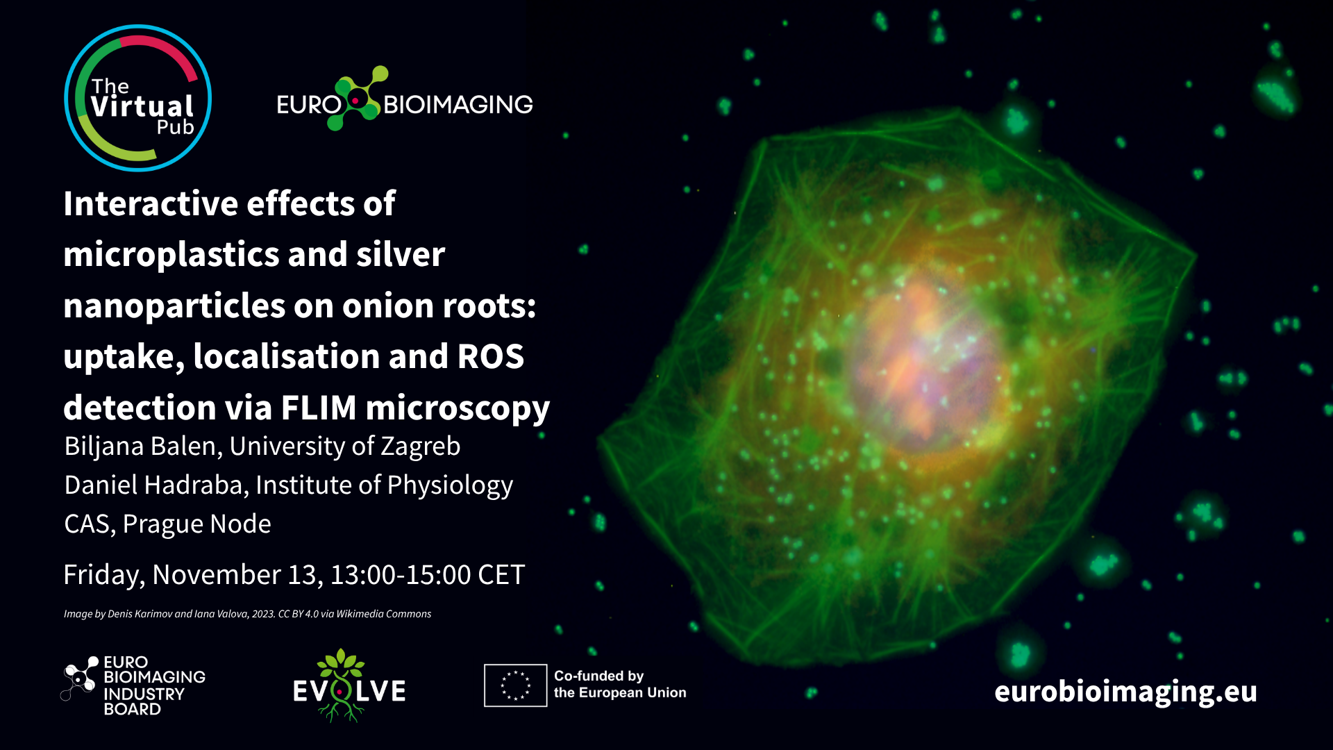
At the Special Edition Virtual Pub, “Imaging Micro- and Nanoplastics,” Euro-BioImaging users, Node experts and Industry partners will explore how advanced microscopy, clinical…
October 16, 2026
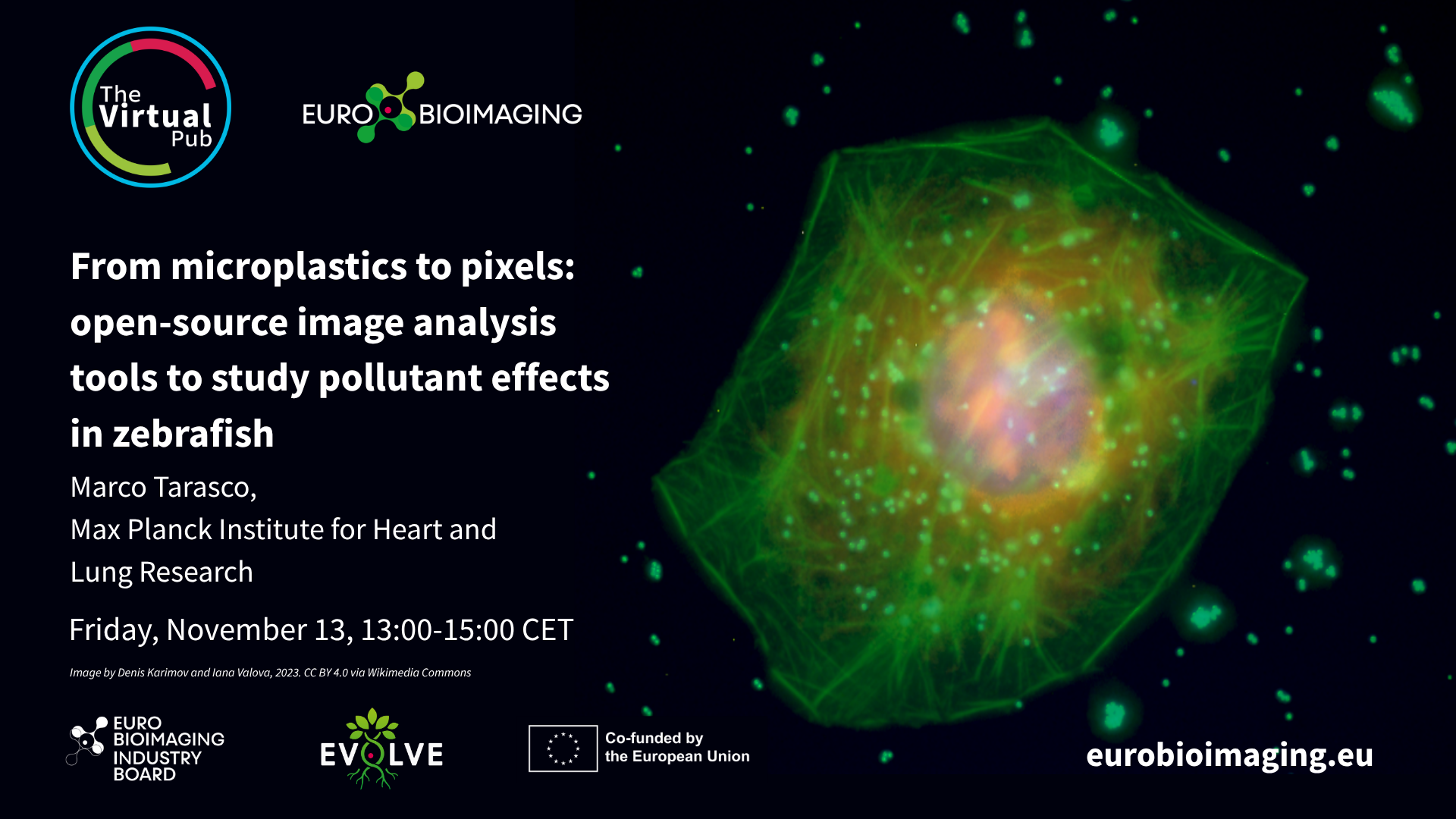
At the Special Edition Virtual Pub, “Imaging Micro- and Nanoplastics,” Euro-BioImaging users, Node experts and Industry partners will explore how advanced microscopy, clinical…