April 28, 2026
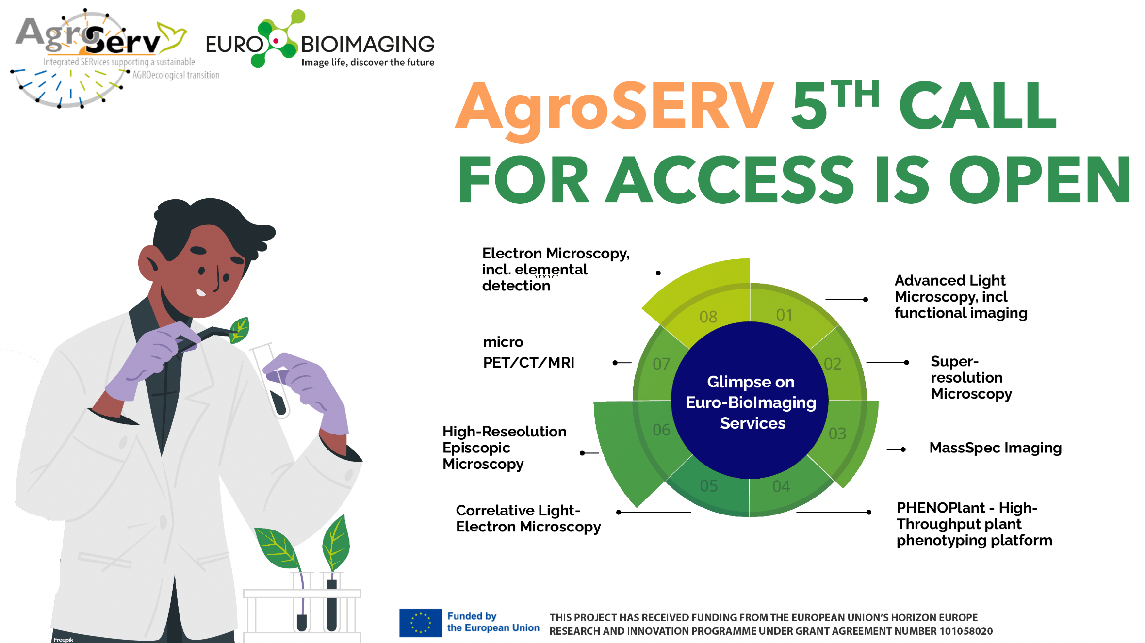
AgroSERV's 5th call for access is open
AgroServ is a transdisciplinary initiative supported by the European Union through the Horizon Europe program and will continue until 2027. It supports the research…

Chiara Cassioli, a Postdoctoral researcher at the University of Siena, is passionate about basic research. Her quest to discover how immune cells communicate within the immune system has been the driving force in her research. So trying to figure out what trafficking pathways are responsible for the biogenesis and the release of SupraMolecular Attack Particles (SMAPs) - a recently discovered way for T cells to kill virally infected and cancerous cells – is right up her alley. And it brought her to the Advanced Light Microscopy Facility at EMBL, part of Euro-BioImaging’s EMBL Node, to carry out a high-throughput, imaging-based RNAi screen project for the first time in her career. We spoke with her to find out more about her fascinating research project and understand why this approach - and the support of the EMBL ALMF - is essential to her work.
Chiara Cassioli got her Master’s degree in Molecular and Cellular Biology from the University of Siena in 2015. She joined the laboratory of Professor Cosima Baldari for her Master’s thesis to study the mechanisms of immune synapse formation. During her PhD research, carried out in the Baldari lab, she had a 6-month stay at the University of Oxford, where she worked in Professor Michael Dustin’s lab. Michael Dustin pioneered the study of immune synapses by developing an innovative imaging approach based on supported lipid bilayers with embedded ligands forming a synthetic antigen presenting cell. Using this technology, Chiara started an in-depth characterization of immune synapses formed by T lymphocytes and optimized a cell line for imaging studies. Later on she even identified a new role for the ciliopathy-related protein Bardet-Biedl syndrome 1 (BBS1) in T cell polarity and immune synapse assembly.
Today, she is back in Professor Baldari’s lab, as a research associate, and she is involved in an exciting ERC Synergy grant project called ATTACK (Analysis of T-cell’s Tactical Arsenal for Cancer Killing). This project brings together researchers from leading labs in the fields of immunology, molecular biology and biophysics to study a previously unknown “weapon” in the T cell’s arsenal, the SMAPs. What makes SMAPs particularly interesting to these researchers is their potential to attack and kill cancer cells in a cell-independent fashion.
SMAPs were first observed by scientists in Michael Dustin’s lab at the University of Oxford, and the paper describing them was published in Science in 2020. While their killing potential has been established, how SMAPs are produced, released and how cancer cells respond to them is as yet unknown. That is the goal of the ATTACK project - to explore this aspect of the T cell cytotoxic machinery and how the results might be translated to new cancer immunotherapies.
Within the ATTACK project, Professor Baldari’s lab is working towards understanding how SMAPs are made inside cytotoxic T cells. To achieve this objective, they will combine two powerful techniques of functional genomics and cell biology, namely RNA interference (RNAi) and high-throughput microscopy, to carry out an imaging-based, high content RNAi screen. They will individually target hundreds of protein-coding genes and will study the impact of each one on SMAP formation. In Siena, Chiara and her team manually acquire images of fixed cells by using a state-of-the-art spinning disc confocal microscope. In Professor Dustin’s lab, Chiara was also trained in Total Internal Reflection Microscopy to study immune synapses. However, the type of work she is doing within the ATTACK project requires ever more powerful imaging capability, beyond the instruments that Chiara has access to in her lab and within her network of partners. That is how she ended up at EMBL’s ALMF, as a Euro-BioImaging user, to work closely with Beate Neumann (aka Faba) to set up a high-throughput RNAi screen aimed at elucidating how the pathways that regulate the trafficking of individual SMAP components converge to allow SMAP biogenesis.
“With a high-content RNAi imaging screen, we can perform 100 experiments in 1,” says Chiara, with stars in her eyes. “At University of Siena, we would have looked at one candidate protein at once, but here, we are using high-throughput microscopy to acquire images automatically from hundreds of samples overnight or overall several days and we are computationally analyzing phenotypes.”
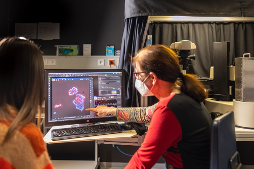
“But we wouldn’t have been able to set up and run this experiment on our own - it’s really a collaboration with the EMBL ALMF. Even before I arrived at EMBL, Faba and the ALMF team had been extremely supportive of our project, advising us at each step of the development of a customer-based RNAi assay, helping us to choose the best cell line and read-out to address our biological question, and helping us to improve our sample preparation to acquire good quality images for co-localization analysis.”
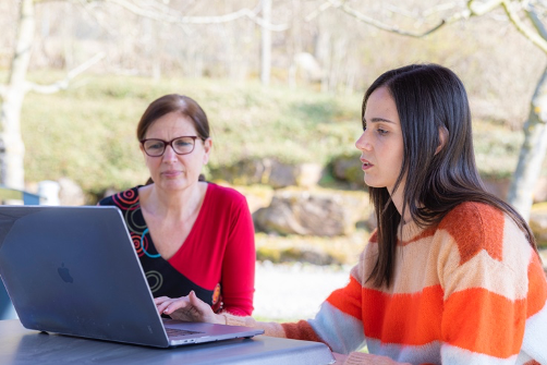
“I feel extremely lucky to have been able to spend three weeks at EMBL’s ALMF as a Euro-BioImaging user. Faba is extremely supportive, and I have learned much from her expertise. She has trained me with several different devices I was not familiar with. And I have her input every time I need it, especially if something unexpected comes up. Every day I discuss with Faba to find the best solution to overcome technical issues and move forward.”
High-throughput RNAi screens are complicated experiments, requiring a lot of support from the imaging facility. Chiara explains the experimental process:
“In our current set-up, we seed the cells on a plate coated with small interference RNAs (siRNA) targeting different genes known or predicted to have a role in the regulation of the secretory pathway and we analyze the phenotype caused by partial depletion or complete removal of the targeted protein by microscopy. We expect to find samples in which SMAP components show different subcellular localizations in the absence of one or more – still unknown – proteins that will be identified as new, potential regulators of SMAP assembly. High-throughput microscopy and computational analysis of phenotypes are obviously crucial to this type of work.”

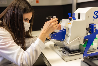
Figure 3: High-throughput RNAi screens are complex experiments requiring support from facility staff. Faba trained Chiara to use several devices she was not familiar with. Chiara coats (left) plates with small interference RNAs (siRNA), on which cells are seeded for solid-phase transfection, fixed and stained (right), then analyzed by high-throughput fluorescence microscopy. Photo copyright: EMBL/PhotoLab Massimo del Prete
“Usually, I apply a hypothesis-based approach – based on prior knowledge and observations I develop a hypothesis as to how a process may work and this hypothesis is then experimentally tested. With high-throughput screening, we will identify new potential candidates involved in SMAP biogenesis through an unbiased approach. The next step will be to validate the most interesting hits and focus on each single regulator for a more in-depth characterization of its function in this process at my home laboratory.”
And this is just what Chiara likes best about her scientific career. Designing and performing experiments, solving scientific puzzles, and discovering something that has never been seen – or explained – before. These are the main reasons that push her to pursue a curiosity-driven career in science.
“I think that people believe having a career in curiosity-driven basic research is not rewarding. But there is so much biology out there – many things we still don’t understand – it is very rewarding to strive for understanding. In this project, I am addressing questions mostly related to unknown SMAP biology. However, in the near future the findings of our research might be rapidly translated into a new SMAP-based cancer-therapy approaches and this makes our project even more rewarding.”
And this is what keeps her going. As she leaves EMBL, she is pleased to have acquired a new skill set and have had a go at a new approach to scientific discovery. And her collaboration with EMBL will continue. There is a large amount of digital image data to sift through, and new cell lines to prepare in the search for understanding SMAP biology.
Chiara would like to thank:
Note: During her visit to Euro-BioImaging’s EMBL Node, Chiara’s travel and lodging expenses were supported by the Italian Fund for Euro-BioImaging User Access, funded by the Italian Ministry of Universities and Research.
More information:
Chiara Cassioli interview in Cell Science:
Cassioli et al. Article in Journal of Cell Science
About the ATTACK Project:
Major ERC-Funding Awarded to Professor Michael Dustin
April 28, 2026
AgroServ is a transdisciplinary initiative supported by the European Union through the Horizon Europe program and will continue until 2027. It supports the research…

April 28, 2026
Since 2024, the EVOLVE project has been empowering Node staff to attend specialized training courses and workshops hosted by fellow Nodes and leading external…

April 27, 2026
Euro-BioImaging is thrilled to announce the introduction of a new imaging technology, Laser Speckle Contrast Imaging, now available in proof-of-concept. This technology can…