Euro-BioImaging is thrilled to announce the introduction of a new imaging technology, Laser Speckle Contrast Imaging, now available in proof-of-concept.
This technology can provide real time visualization of local microcirculation to investigate dermal perfusion, skin inflammation, wound healing, as well as acute and chronic inflammation joint disorders. Beyond dermatological and inflammatory applications, Laser Speckle Contrast Imaging is also relevant for monitoring cerebral blood flow, investigating stroke and ischemic models and conducting functional brain activation studies.
We met Dr. Zsófia Hajna, from the University of Pecs, part of the Medical and Preclinical Imaging Hungarian Node, to get more insight about the technology and its main application fields.
Zsófia is a senior lecturer and part of the staff working at the Preclinical Imaging Platform at Pécs University. She works at the Department of Pharmacology and Pharmacotherapy (Medical School, University of Pécs) under the supervision of Dr. Zsuzsanna Helyes and she is the specialist responsible for Laser Speckle Imaging.
Zsófia, could you briefly introduce Laser Speckle Contrast Imaging and explain its scientific relevance? When did this technology begin to play an important role in preclinical research?
Laser Speckle Contrast Imaging is a non-invasive, contactless and fast imaging method that is highly suitable for wide-field visualization and real-time monitoring of blood perfusion in various inflammatory and degenerative conditions without using exogenous contrast media. This technique is based on the principle that if a coherent laser light illuminates a biological tissue, the backscattered light produces a random interference pattern, called speckle pattern. If there are moving particles in the tissue – such as blood cells –, their movement will cause fluctuations in the speckle pattern, strongly correlating with the intensity of microcirculation.
Laser Speckle Contrast Imaging was introduced in the early 1980’s, when Fercher and Briers reported the assessment of retinal capillary blood flow as the first biomedical application of this method. At that time, acquisition and processing of the speckle pattern images were performed via simple, non-digital photography, enabling only qualitative investigation of blood perfusion. Later, in the 1990s, digital image processing of the speckle photographs was developed providing real-time mapping of the measurement area and semi-quantitative assessment of tissue microcirculation. In the last 20-25 years, both the preclinical and clinical use of Laser Speckle Contrast Imaging has rapidly spread, with continuously broadening application fields and parallelly developing techniques (e.g. quantitative approach).
What are the principal research areas in which it can be applied?
In the preclinical research, Laser Speckle Imaging can be well applied for the investigation of dermal perfusion and skin inflammation, such as the models of cutaneous neurogenic inflammation, allergic contact dermatitis and psoriasis. Further application fields are acute and chronic joint inflammation, as well as various forms of ischemic and neuropathic states of the extremities The process of wound healing can also be well visualized under postoperative condition or after burn injury. Besides, the investigation of meningeal microcirculation is particularly informative in modelling migraine.
In clinical research, Laser Speckle is well applicable in the investigation of rheumatological diseases, as well as systemic sclerosis. This imaging technique can also be used for the diagnosis of peripheral arterial disease and peripheral neuropathy, as well as the assessment of burns and wound healing. Furthermore, several dermatological disorders, such as allergic skin reactions can be determined.
Can Laser Speckle Contrast Imaging be integrated with other imaging technologies available at your institute? If so, how do these combinations enhance research outcomes?
Inflammatory and degenerative processes usually lead to alterations of microcirculation. However, further symptoms such as plasma extravasation, vascular disruption, as well as the accumulation and activation of white blood cells, or even structural remodelling also often occur. Therefore, it is highly essential to visualize these changes as well, and for this purpose, several other imaging techniques are available in our laboratory. For example, bioluminescent and fluorescent signals of the inflammatory processes can be well visualized with IVIS Lumina II in vivo imaging system and fluorescent molecular tomography, and for the investigation of structural alterations micro-CT analysis is also available. These methods can be parallelly incorporated in the same project, thus enhancing the complexity of the study and strongly contributing to the detailed and thorough investigation of various inflammatory and degenerative conditions.
What are the main advantages of this technique?
First of all, Laser Speckle Contrast Imaging offers a non-invasive and non-contact approach of blood perfusion investigation. Moreover, there is no necessity of chemical tracers or dyes, making both the preclinical and the clinical measurements simple and user-friendly. Full-field visualization of the microcirculation can be performed via high resolution color-coded images providing illustrative figures based on the settings adjusted to the researcher’s and the project’s requirements. Furthermore, the high speed of the measurement also enables the determination of real-time graphs, promoting the dynamic assessment of vascular responses. Therefore, this technique provides adequate visualisation and high-quality assessment of tissue microcirculation.
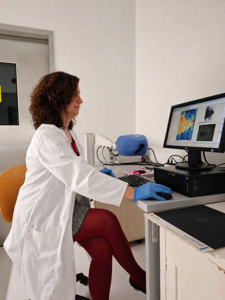
Dr. Zsófia Hajna, from the University of Pecs, part of the Medical and Preclinical Imaging Hungarian Node, performing Laser Speckle Contrast Imaging.
More news from Euro-BioImaging
July 23, 2026
Cellular Imaging Hungary Node expands expertise in advanced neurophotonics
The Euro-BioImaging Cellular Imaging Hungary Node has expanded its service portfolio with the addition of the BrainVisionCenter (BVC) in Budapest. Following a successful…
The UK Euro-BioImaging Node expands from seven to thirteen sites!
We’re delighted to announce that the UK Euro-BioImaging Node is expanding, growing from seven to thirteen sites following a successful upgrade application and…
Building Skills Across Europe: EVOLVE Supports the Second Edition of the Distributed Image Analysis Training Course
The second edition of the “Introduction to Image Analysis with Python for Life Scientists” course marked another successful milestone in the development of Euro-BioImaging’s distributed training model.