May 27, 2025
Euro-BioImaging Board Meeting in Amsterdam
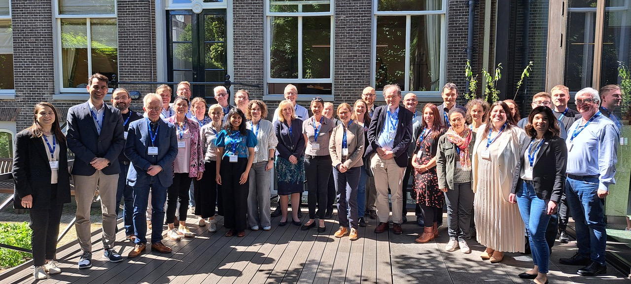
The 12th Euro-BioImaging Board meeting took place in Amsterdam, from May 13-14, 2025, hosted by our colleagues from NL-BioImaging. It was the occasion for…
May 27, 2025
The 12th Euro-BioImaging Board meeting took place in Amsterdam, from May 13-14, 2025, hosted by our colleagues from NL-BioImaging. It was the occasion for…
March 12, 2025
The EVOLVE Job Shadowing program is a fantastic opportunity for Node staff to immerse in the daily life and operations of other Nodes of…
February 12, 2025
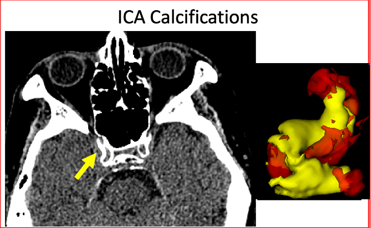
What could be more fascinating than to explore the prevalence of cardiovascular disease in populations as diverse as ancient mummies, modern Americans, and people…

October 21, 2024
Euro-BioImaging is thrilled to be part of ILLUMINATE (Increasing Lutetium production while leveraging metabolic imaging to enhance theranostics effectiveness), a pioneering 54-month public-private partnership…

June 20, 2024
The 2024 Annual Congress of the European Association for Cancer Research (EACR 2024) took place in Rotterdam from June 10 to 13, 2024. The…

July 23, 2023
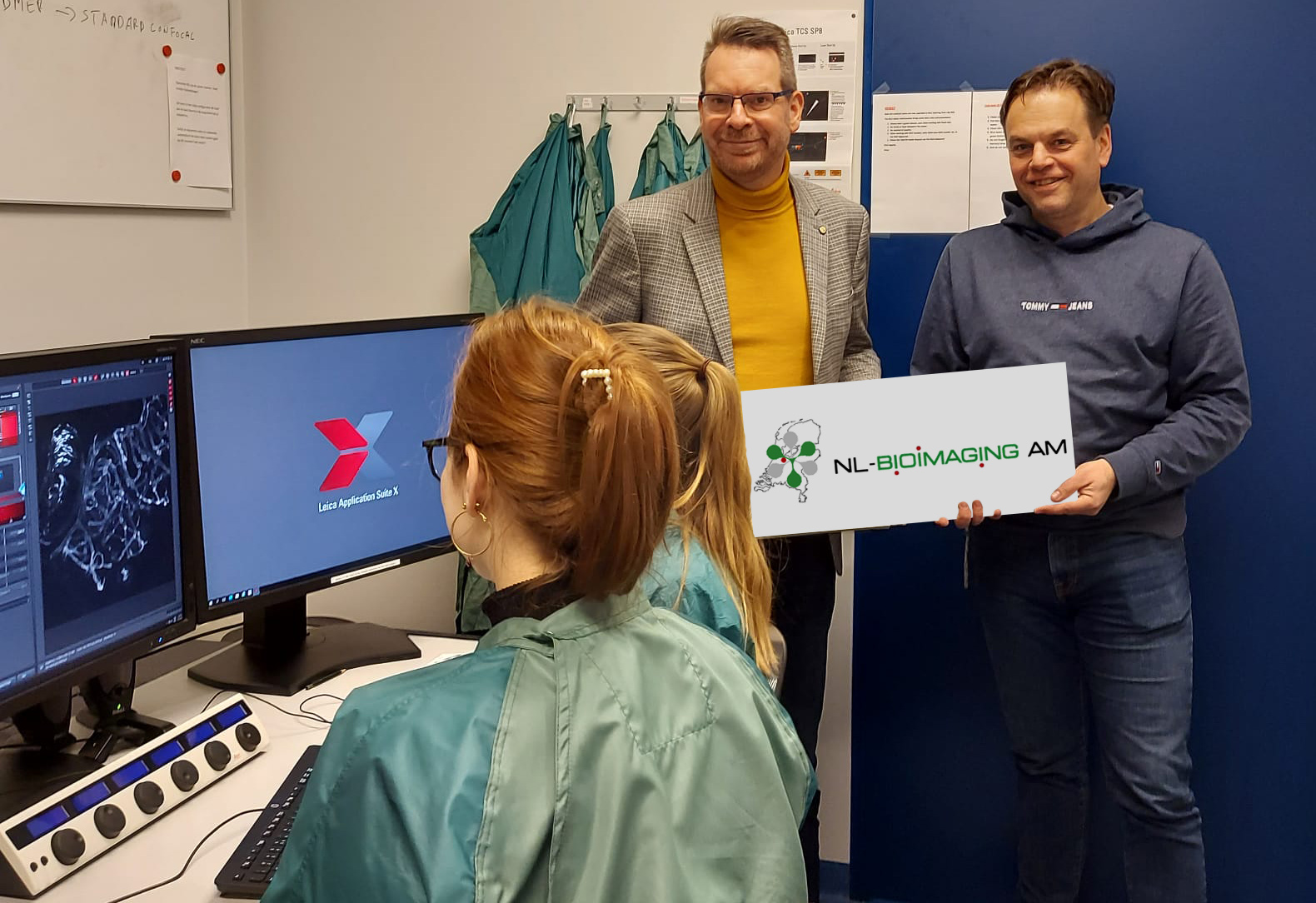
What is Euro-BioImaging? What is open access to imaging? As part of a series spotlighting Euro-BioImaging Nodes, we asked Marc van Zandvoort, Chair of…

June 19, 2023
This year, the European Light Microscopy Initiative meeting took place in Noodwijkerhout, the Netherlands, organised by NL-BioImaging. Several Hub team members were able to…
February 20, 2023
Three hundred years after Antoni van Leeuwenhoek, father of live-cell microscopy, performed his last amazing research, a 25 Million Euro grant from the…
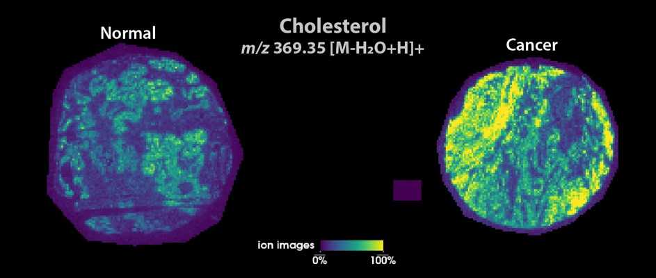
September 1, 2022
How does prostate cancer become aggressive and life-threating? That is a question that Maria K. Andersen, post-doc researcher at the…

July 19, 2022
One of Euro-BioImaging’s key missions is to facilitate excellent science by providing open access to imaging technologies and expertise. Users come from many backgrounds…

May 24, 2022
The Challenges Framework flagship Node organizes a series of Grand Challenges in Medical Image Analysis (www.grand-challenge.org), focused at standardized evaluation…
March 21, 2022
In 2021, Euro-BioImaging launched its pilot call for funded user projects to be supported by an internal Euro-BioImaging User Access Fund.