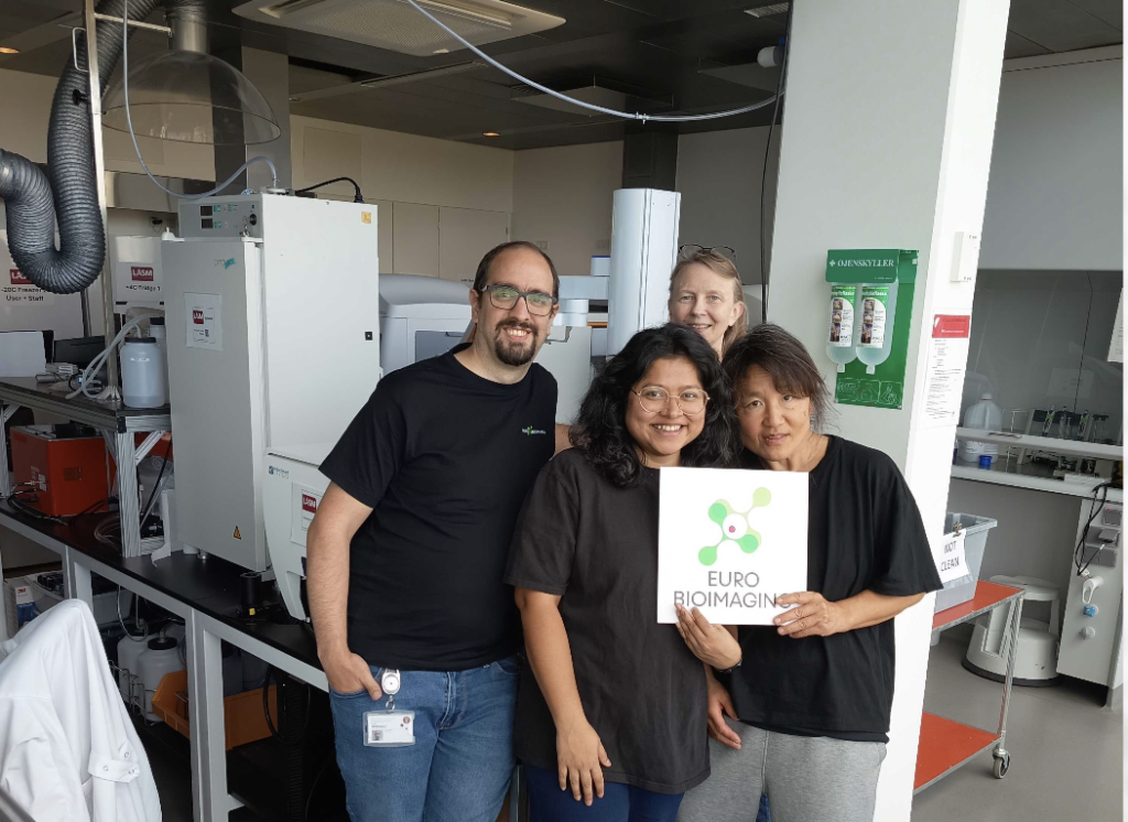
We are delighted to welcome the Laboratory, Automation, Screening, and Microscopy facility at the Biotech Research & Innovation Centre (BRIC), University of Copenhagen, to the Euro-BioImaging family. This facility brings a unique range of expertise to the Euro-BioImaging technology portfolio, including Light Microscopy and High-Content CRISPR Screens. Because of their unique expertise, their application was highly recommended by the Euro-BioImaging Scientific Advisory Board in the recent Node upgrades. We look forward to working with our new colleagues!
LASM is a newly established core facility originating from the merging of the Light Microscopy and High-Content CRISPR Screens facilities within Biotech Research and Innovation Centre (BRIC), University of Copenhagen. The new facility has staff members specializing in supporting users and in different roles: screen design, automation, microscopy and image analysis.
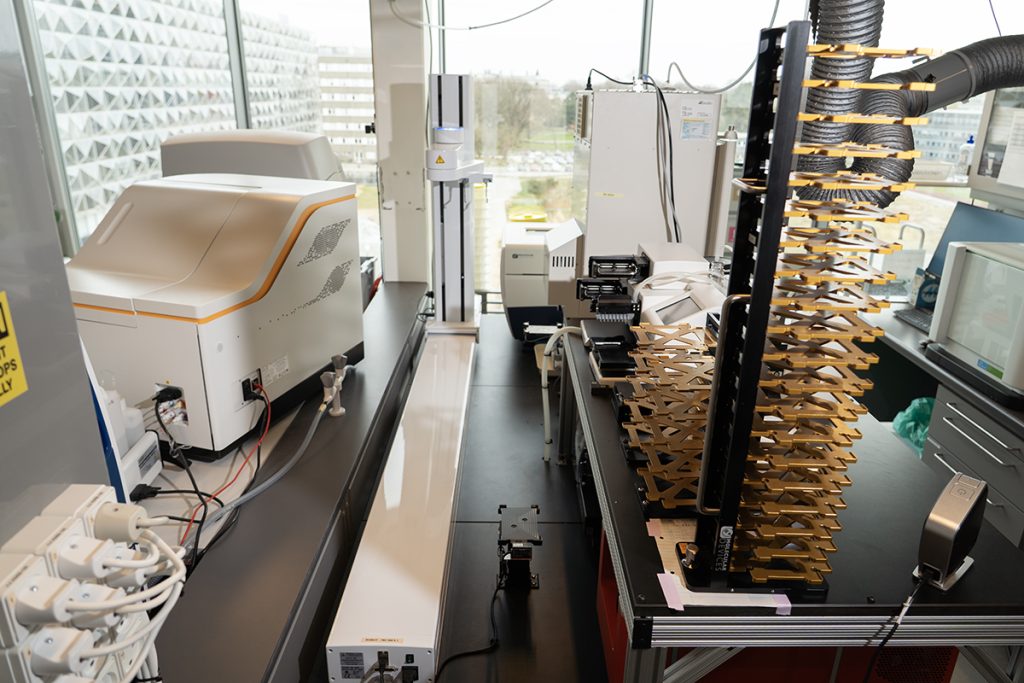
LASM offers standard microscopes for daily use, but its focus is on high content microscopy and screening. LASM offers several high-throughput widefield microscopes for live-cell images (1x Evos FL Auto, 2x Olympus ScanR, and 1x Incucyte S3), a high-throughput spinning disk confocal (ImageXpress HT.AI) and a multiplexing microscope (MACSima).
In addition to the microscopes, LASM possesses an automation platform to support arrayed screening. This includes a mix of acoustic dispensers, peristaltic pumps, and pipetting robots, allowing reproducible preparation of assay plates for analysis in the high-throughput microscopes.
"We're really happy to join the Euro-Bioimaging community. It's such a great opportunity to exchange with other facilities and to share our services with an even larger group of users, no matter if they come from industry or academia."
-- André Dias
The automation platform can be used to support both experimental setup and staining procedures. This allows for a reduction of the amount of sample and reagents used by enabling transfer of nanoliter sized volumes, which can be especially important for working with expensive reagents or limited size samples (e.g. patient material).
The BRIC facility comprises an automation platform to support arrayed screening.
LASM offers a cherry pickable drug library, as well as two whole genome human sgRNA libraries (CRISPR knock-out and activation) enabling users to select a limited panel of genes of interest to their studies. The staff is also available to help with experiment design so users can get the most out of their experiments.
Resources (both computers and software) and support for data analysis are available for both local and remote access. This includes both proprietary software from the microscope manufacturer and open-source software developed by the community, as there is a dedicated staff member intended to assist users with image and data analysis.
The facility is fully open to researchers in academia as well as external and industrial researchers.
About Danish BioImaging Node- Euro-BioImaging As a Life Science researcher, you have open access to high end equipment and expertise at the BioImaging Core Facility at the Aarhus University, the preclinical medicine imaging facility at Aarhus University Hospital, the Danish Molecular Bioimedical Imaging Center (DaMBIC) at the Syddansk Universitet – University of Southern Denmark, the Center for Advanced BioImaging (CAB), the Core Facility of Integrated BioImaging (CFIB) – University of Copenhagen and the DBI INFRA Image Analysis Core Facility.
More news from Euro-BioImaging
October 23, 2026
Plastic pollution and health: a preclinical PET FDG study
At the Special Edition Virtual Pub, “Imaging Micro- and Nanoplastics,” Euro-BioImaging users, Node experts and Industry partners will explore how advanced microscopy, clinical…