
October 15, 2026
Open Call: Exploit Large Scientific Datasets with AI and HPC through RI-SCALE
The European RI-SCALE project has launched an Open Call for Use Cases, inviting research teams, research infrastructures and companies to…



The Electron Microscopy Unit (EMBI) provides biological electron microscopy services at the University of Helsinki. Led by Research Director Eija Jokitalo, this facility is also part of Euro-BioImaging’s Finnish Advanced Microscopy Node (FiAM). As of December 2025, it became the first imaging core facility in Finland to join ZEISS’s exclusive “labs@location” network, a network of leading research facilities with an excellent reputation for implementing technologies, applications, and workflows. We spoke to Eija Jokitalo and Toni Montonen, Account Manager, Research Microscopy Solutions (RMS), ZEISS Nordics, to learn more.
“The EMBI facility is unique in Finland,” says Toni Montonen. “Their expertise in biological Electron Microscopy (EM) is recognised across Europe. It’s a world class facility - with the whole pipeline from sample preparation to imaging to image processing and visualisation. They have developed expertise that we don’t have at ZEISS - which is why I nominated them to become a labs@location site,” explains Toni.
Becoming a labs@location site is an honor. Each facility goes through a stringent international selection process. It’s a stamp of approval, a recognition of the facility’s expertise. It’s also an opportunity to join a worldwide network of high-end labs and core facilities that meet regularly to exchange knowledge. And it’s a responsibility. ZEISS trusts these facilities to demo their instruments to potential customers, leveraging their expertise to show other customers the possibilities of a system.
“I see several advantages in having a close collaboration with ZEISS,” says Eija Jokitalo. “First, the proximity with the company allows me to learn where the field is going. The first time I heard about Serial Block Face Imaging was from a company representative. It was in the early days of development – a few years before the first commercial system came out. By learning about the new system early on, I had time to negotiate within my consortium and request funding. Thanks to this insider knowledge, we became an early adopter of the technology and were able to obtain one of the first 10 commercial systems in the world. Now, as part of the labs@location network, I believe we will have even more information about where the field is going.”
“Secondly, it’s a way for us to give feedback and influence developments. We know the challenges our users encounter, and understand the questions they want to address,” states Eija. “This can be of interest to ZEISS. And finally, networking is really important to me. I look forward to meeting with the labs@location network, identifying collaboration areas, and potentially developing workflows together.”
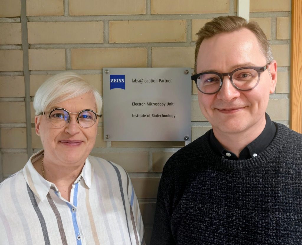
“Indeed, labs@location is designed as a two-way street. Both sides have a lot to gain from this collaboration,” confirms Toni.
With only two ZEISS systems, EMBI is one of the smaller facilities to join the labs@location network, attesting to their strong expertise. Their Crossbeam 550 FIB-SEM was installed during COVID-19 and the Gemini 460 with Volutome was installed in summer 2025. Their labs@location nomination was approved in August 2025.
With so many things to celebrate, Eija and her team organised a Symposium entitled “Frontiers in Imaging: Celebrating the EMBI-ZEISS partnership” in Helsinki in December 2025. This event recognized Eija’s staff for the excellent work they do in the field, inaugurated the two Electron Microscopy systems installed in the facility, and celebrated the role of EMBI in the Volume EM community. The programme featured an opening address from John Eriksson, Director General of Euro-BioImaging, who played a big role in building the Finnish national imaging community, and a keynote address on “Multimodal correlations allow precise targeting of volume Electron Microscopy in complex model and non-model organisms” by Yannick Schwab, Head of the Electron Microscopy Facility at EMBL, a close colleague within the Volume EM community. There were also speakers from ZEISS, and three of EMBI’s users shared the amazing research they are undertaking in plant science, tissue studies, and cell biology, nicely demonstrating the range of possible applications.
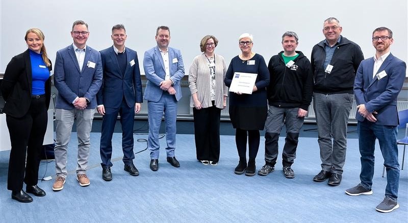
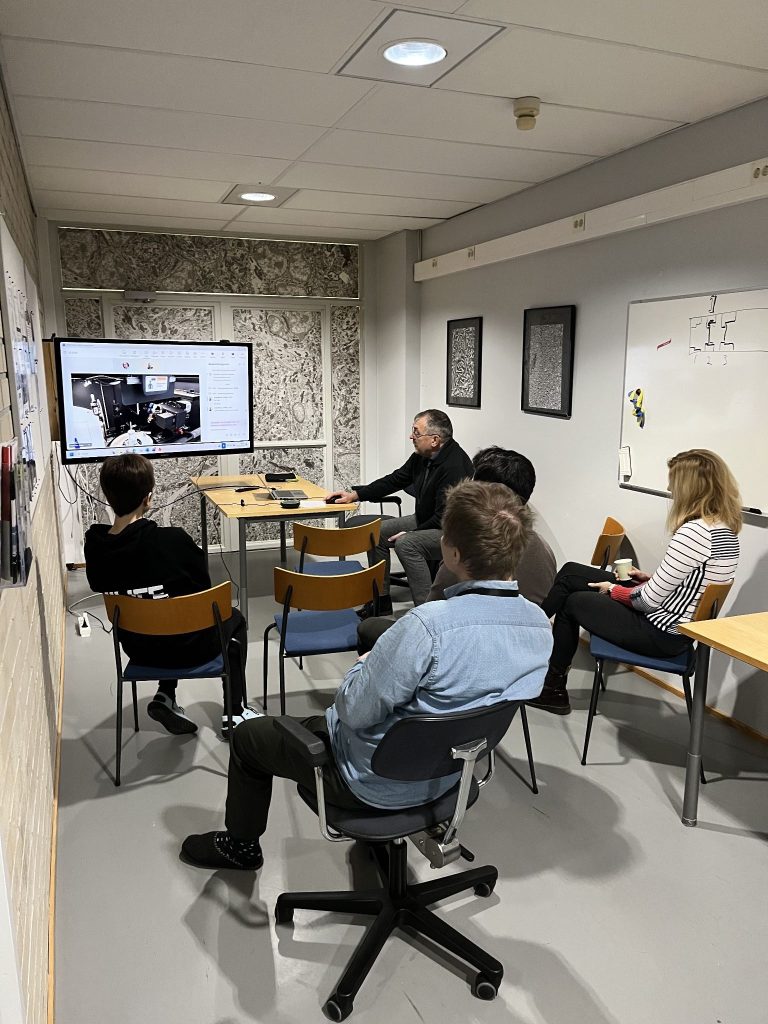
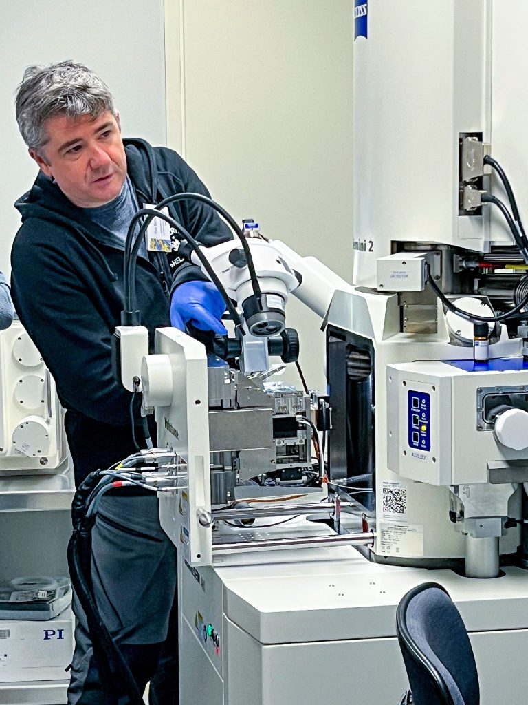
There were also demos – Eija’s team showed how the volume EM microscopes can be used, and a specialist from Zeiss gave a workshop on image analysis. A highlight of the symposium – and a perfect illustration of Eija’s point about access to insider knowledge – was the XRM Micro CT web demo – via a virtual connection to the ZEISS site in Oberkochen.
“XRM imaging is currently available at very few places in Finland,” explains Toni. “So the demo was something special. I think it helped raise awareness of this new technology, and its potential for correlated workflows.”
The EMBI facility is strategically positioned as Finland’s state-of-the-art for Volume EM – and it is an excellent place to carry out a collaborative imaging project, with the support of the expert team. Via Euro-BioImaging, EMBI offers open access to its technologies and expertise to all researchers, regardless of their career stage, affiliation or scientific area.
The heart of EMBI’s mission remains user access and user support. Under Eija’s lead, they seek to keep their competitive advantage. “If you want to be in the forefront, and an active player, if you want to provide the latest, coolest technologies to the users, you need to know where the field is going. If you are learning about technologies by reading published papers, it’s too late.” Being a labs@location facility is one important way for them to do so.
Congratulations to EMBI for this important recognition of your expertise and stellar example of industry-academia partnership!
October 15, 2026
The European RI-SCALE project has launched an Open Call for Use Cases, inviting research teams, research infrastructures and companies to…

October 15, 2026
The 2026 edition of SPAOM, the meeting of the Spanish and Portuguese Advanced Optical Microscopy community, marked a special milestone: the 10th edition of this meeting bringing together researchers, microscopy specialists, technology developers and imaging facilities.

October 13, 2026
From 27 September to 1 October, our team member Feriel Ramdhane (Image Data Scientist at Med-Hub) represented Euro-BioImaging at MICCAI 2026 in Strasbourg.