New imaging technologies are developed at a rapid pace and keeping up with these advancements is a key mission for Euro-BioImaging. At the same time, we have an important responsibility to our users, to ensure that new technologies are service-ready for open access use before being included in the Euro-BioImaging portfolio. To balance the need for fast availability of new technology with the requirement for due diligence to ensure service-readiness, Euro-BioImaging implements a defined process. New technologies of interest are constantly identified by the community, pass initial evaluation of the service model by the Euro-BioImaging Hub, and are then tested at our Nodes in open access service provision. After this two-step process, called Showcasing and Proof-of-Concept, the technologies are evaluated by our Scientific Advisory Board. If the evaluation is successful, the addition of the technologies to the Euro-BioImaging portfolio can be approved by the Euro-BioImaging Board.
Since the start of operations in 2019, several technologies have been showcased at Euro-BioImaging. In the first round, these were all established technologies, showcased to fill in gaps in the technology portfolio. The Proof-of-concept study for these technologies started in September 2020, making them available in open access "under probation" at several Euro-BioImaging Nodes. Concluding the proof-of-concept study was an evaluation by our Scientific Advisory Board in Fall 2022, taking into account feedback from users and Nodes, the access model, and user demand.
We are pleased to announce that all four technologies successfully passed the proof-of-concept study and were unanimously recommended to be included within the Euro-BioImaging technology portfolio by our Scientific Advisory Board. They were subsequently officially approved by the Euro-BioImaging Board in November 2022. Now these four technologies join our official technology portfolio and continue to be available in open access via various Euro-BioImaging Nodes. The approved technologies are SIM, AFM, QPI and MSI .
With the official Board approval the proof-of-concept study process has now been fully completed for the first set of technologies, highlighting the practical functionality of the established processes at Euro-BioImaging to expand its technology portfolio. The process will be applied in the same way to bring on board and assess the technologies currently still in proof-of-concept studies and those new technologies that will enter into proof-of-concept study status in January 2023.
We wish to thank all Nodes who participated in this successful first proof-of-concept as well as the Scientific Advisory Board for their participation in the evaluation process.
Learn more:
QPI
Are you working on drug testing, cell interactions or cancer cell biology? Try out Quantitative Phase Imaging!
Helena Chmelová, Ph.D., from Light Microscopy Core facility at Institute of Molecular Genetics, Prague, Czech Republic, part of Euro-BioImaging’s Advanced Light and Electron Microscopy Node Prague, explains how Quantitative Phase Imaging (QPI) is very useful for studying cell shape, viability, motility, polarity, growth or differentiation under various conditions. Learn more here
SIM
Would you like to study intracellular structures in super resolution? Try out Structured Illumination Microscopy (SIM)!
Ivan Novotný, Ph.D., from Light Microscopy Core facility at Institute of Molecular Genetics, in Prague, introduces us to Structured illumination microscopy (SIM) imaging technology to visualize intracellular structures. Learn more about how it compares to other super-resolution techniques, and what applications it can be used for:
Interested in the spatial distribution of biomolecules from cellular and tissue surfaces at the nm and µm scale? Then Mass-Spectrometry-based Imaging (MSI) could be for you!
The Facility of Multimodal Imaging – AMMI Maastricht Node offers a unique infrastructure for state-of-the-art mass spectrometry-based molecular imaging (MSI). This technology can be used to study tumour heterogeneity at the molecular level, to study the distribution of metabolites, proteins, lipids or glycans in a particular tissue specimen but also to follow the biodistribution of a drug for PK&PD.
Learn more in this interview with Dr. Berta Cillero Pastor:
Spatial distribution of cholesterol on a prostatic normal (left) and a cancer (right) sample. Image courtesy of Maria K. Andersen.
How can MSI support prostate cancer research?
Learn more in this interview with Euro-BioImaging user Maria K. Andersen, post-doc researcher at the Norwegian Institute of Science & Technology (NTNU) – Trondheim.
Want to analyze metabolite or lipid distribution across tissue sections? Why not try out Mass-Spectrometry-based Imaging (MSI) for biological applications?
The power of MSI is that it enables us to perform studies of metabolomics, proteomics and lipidomics without needing to tag the molecules of interest. Our expert, Seetharaman Parashuraman, Head of the Bioimaging facility of the Institute of Biochemistry and Cell Biology, Naples, an institute belonging to the National Research Council, part of our Advanced Light Microscopy Italian Node, gives some hints on sample preparation and explains the advantages of this technology.
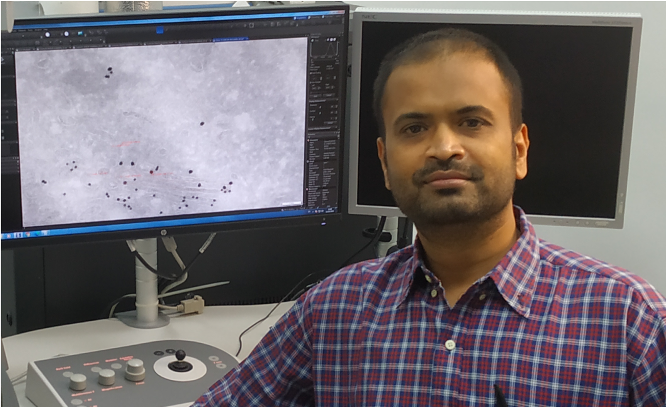
Working with membranes? Need a technology with vertical and lateral resolution in the nanometer range and requiring only a small number of acquisitions? Try Atomic Force Microscopy!
Atomic Force Microscopy (AFM) is a technique allowing the characterization of sample morphology at nanoscale. Images are acquired by raster scanning a nanometric tip in gentle contact or intermittent contact with the sample. By measuring the tip-sample interaction force as a function of the tip-sample distance, AFMs can evaluate sample elastic and viscous properties. Learn more in this interview with Camilo Guzmán, manager of the Finnish Advanced Light Microscopy Node
Camilo Guzmán, manager of the Finnish Advanced Light Microscopy Node explains that AFM is one of the most suitable instruments for experiments where molecular binding forces need to be measured or where the mechanical properties of a material need to be characterised at a micrometric scale and under very low forces (pN to nN). AFM not only is very precise, but it is highly versatile to different materials and samples and furthermore it can be combined with fluorescence microscopy to give a full description of the sample that is being tested.
More news from Euro-BioImaging
July 24, 2026
A Multiplexed Vision: Highlights from the Special Edition Virtual Pub on Spatial Transcriptomics
On June 12th, Euro-BioImaging hosted its latest Special Edition Virtual Pub, bringing together over 200 researchers and technology developers working at the cutting…
Cellular Imaging Hungary Node expands expertise in advanced neurophotonics
The Euro-BioImaging Cellular Imaging Hungary Node has expanded its service portfolio with the addition of the BrainVisionCenter (BVC) in Budapest. Following a successful…
The UK Euro-BioImaging Node expands from seven to thirteen sites!
We’re delighted to announce that the UK Euro-BioImaging Node is expanding, growing from seven to thirteen sites following a successful upgrade application and…