
March 26, 2026
Welcome, MAdMiC Node!
The Madrid Advanced Microscopy Center (MAdMiC) is the first Euro-BioImaging Node in Madrid (Spain). It is formed through the collaboration and close work of…

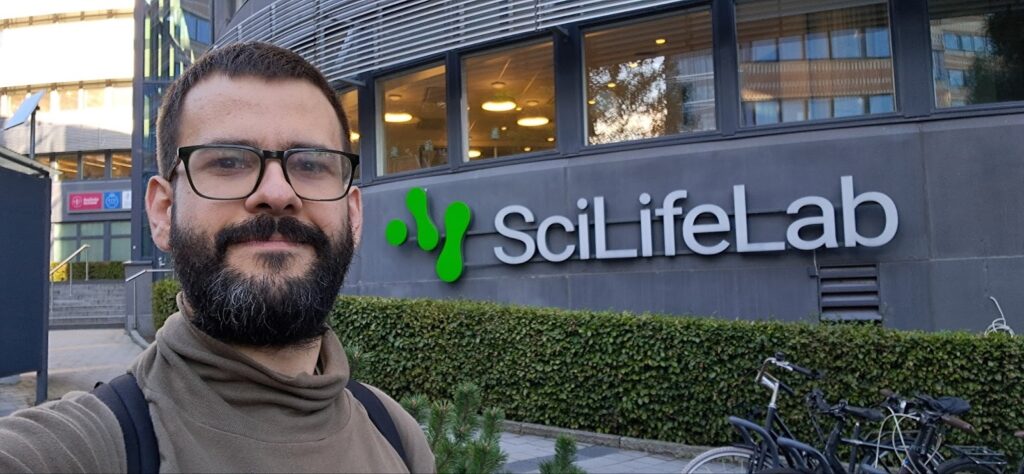
Diego Ramirez is a microscopy specialist helping users with optical microscopy techniques at the Biophysika Unit, part of the Euro-BioImaging’ Bilbao Node. Ana Agostinho is a super-resolution microscopy specialist at SciLife Lab’s Advanced Light Microscopy facility in Stockholm, part of Euro-BioImaging’s Swedish NMI Node. Their facilities are located 2,800 kms away from each other, yet their paths crossed as part of the Euro-BioImaging/EVOLVE job shadowing programme, an innovative initiative designed to increase exchanges between Euro-BioImaging Node staff. In September 2025, Diego spent four days in Stockholm shadowing Ana, to optimise his Expansion Microscopy technique. A great experience for both, and one which has already enriched the service offer of the Bilbao Node.
Expansion Microscopy (ExM) is a growing technique, making it possible to obtain super-resolution results without acquiring new or specific equipment. Without going into too much detail, the method involves crosslinking molecular targets in a fixed sample to a swellable polymer, which can be physically expanded by immersion in distilled water. (Learn more here) It allows scientists to look at subcellular structures such as mitochondria, centrioles and microtubules in super-resolution using a conventional, diffraction-limited microscope.
At the Bilbao Node, where Diego has been working since early 2025, the team uses several Expansion Microscopy protocols, but weren’t thrilled with their results. When Diego heard about the EVOLVE job shadowing programme, he saw it as an opportunity to benefit from the vast experience of other Node staff within Euro-BioImaging, to learn more about ExM.
Ana with her colleague Steven Edwards have been offering ExM as a service in the facility since 2021. Together, they help 4-5 users each year with this technique. A recent publication in EMBO Journal entitled “Spatial mapping of DNA synthesis reveals dynamics and geometry of human replication nanostructures” demonstrates the power of this technique for the detection of nuclear nanostructures and demonstrates how the facility supports users.
On top of that, Ana and Steven are really happy to help other core facility staff master the technique. They’ve even developed a three-day hands-on programme to share their knowledge of expansion microscopy with core facility staff, and tested it with several visiting facility staff.
“The Euro-BioImaging/EVOLVE job shadowing visit really fit my expectations. I got lots of training on how to prepare gels, how to visualise the samples….Together, we explored the best way to apply the protocols with the resources we have [at my home institute]. It was great to get hands on experience, and advice on how to improve results with our set-up in Bilbao.”
-- Diego Ramirez, Biophysika Unit, Bilbao
“We really enjoy working with other core facility staff, and sharing our insights with them. Working with gels is really tricky, and developing a protocol that works for your sample can be difficult. We’ve tried a lot of things and can help with trouble-shooting, and discuss some tips and tricks to help the users feel comfortable performing this technique back in their labs or research facilities.” says Ana Agostinho.
Swedish NMI was a perfect fit for Diego’s job shadowing visit, not only because of their extensive ExM expertise (and experience teaching it), but also because of the availability of MINFLUX, a technology Diego was curious about.

“The Euro-BioImaging/EVOLVE job shadowing visit really fit my expectations,” says Diego. “Throughout my stay, Ana was super supportive. Starting on Day 1, I got to work with the gels. I got lots of training on how to prepare gels, how to visualise the samples….and we took time to discuss the data. She also listened to me when I explained the situation in my home facility. Together, we explored the best way to apply the protocols with the resources we have there. It was great to get hands on experience, and advice on how to improve results with our set-up in Bilbao.”
“ExM isn’t just one protocol; it’s a lot of different protocols. Ana and her colleagues have tried many of them,” says Diego. “So I got lots of tips from these experts!!”
Diego also appreciated exploring other super resolution techniques at Ana’s facility. “My three-day visit offered additional perspective regarding expansion microscopy implementation and general knowledge in MINFLUX microscopy that allowed us to have a more critical view of how it works and the value that can provide us,” affirms Diego.
“We really enjoy working with other core facility staff, and sharing our insights with them. Working with gels is really tricky, and developing a protocol that works for your sample can be difficult. We’ve tried a lot of things and can help with trouble-shooting, and discuss some tips and tricks to help the users feel comfortable performing this technique back in their labs or research facilities.”
-- Ana Agostinho, SciLifeLab ALM Facility, Stockholm
After coming back from Sweden, Diego immediately implemented everything he learned. After the first week, his results improved dramatically. He increased the signal a lot, and reached a much higher quality with the images. He was able to identify what could be improved, correct what he had been doing wrong, and even think about redesigning the workspace, to make it more efficient. “All of these steps helped me to improve my protocol, raise my capacity, and process many more samples,” says Diego.
Many of the users of the Bilbao Node work with culture cells, including nuclear pores, predominantly for basic research. Using ExM to look at brain tissue samples will be an area to explore in the near future, in particular for neuronal connections, to get a much better idea how the receptors in the synapses work. All of the knowledge Diego gained during the job shadowing visit will be shared with the rest of the researchers, technicians and students at the Bilbao Node, an important step to raise interest and promote the use of expansion microscopy.
In conclusion, Diego reflects on the impact of the Euro-BioImaging/EVOLVE job shadowing programme. “It was a very positive experience that allowed us to learn from each other, to discover new things that can be both unknown or not previously taken into account. This is possible because of different research focus and thinking styles. The Job shadowing program allows both partners to get mutual knowledge from each other, as well as to accelerate and improve successfully the implementation of a technique in another Euro-BioImaging Node.”
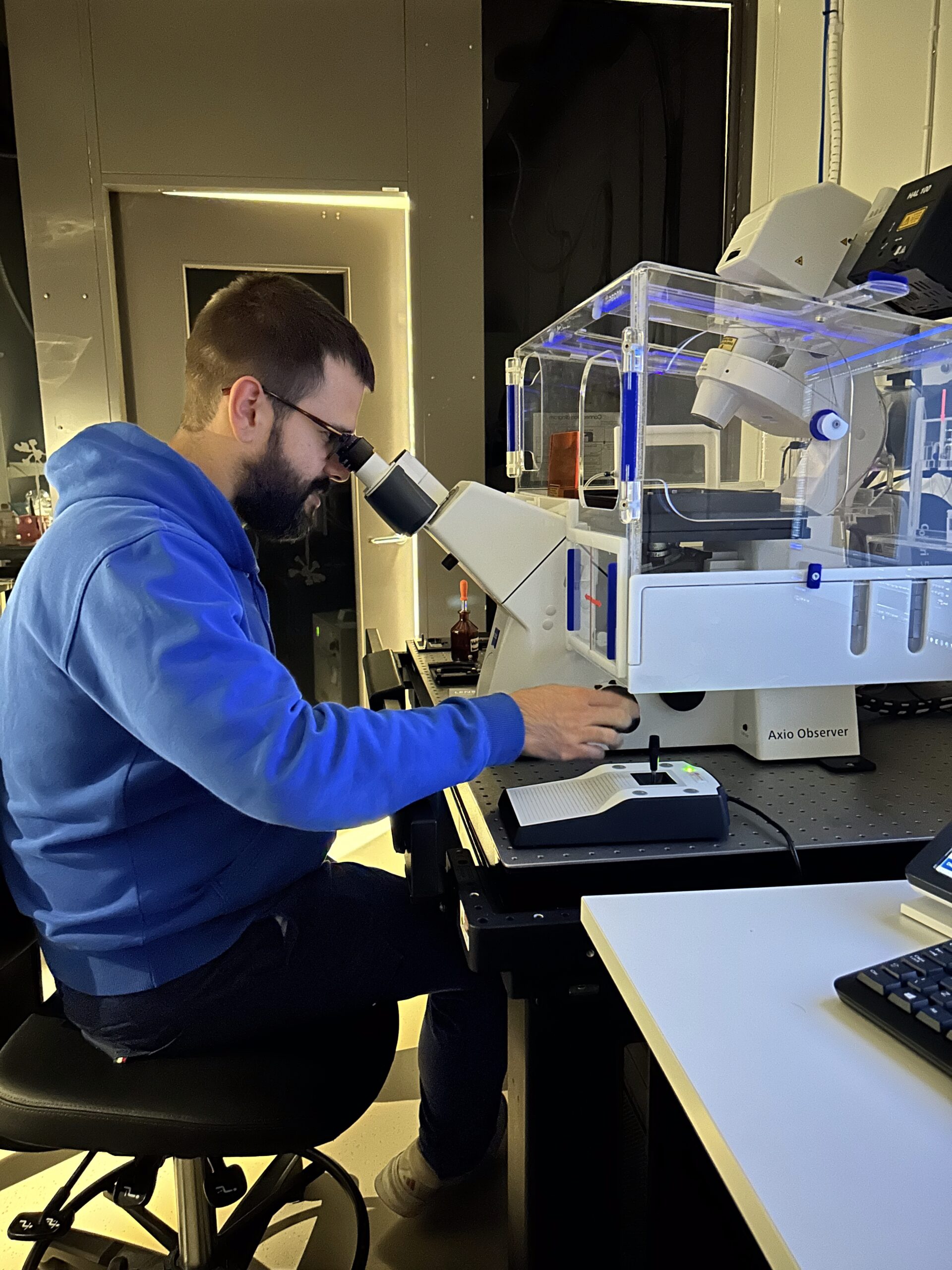
Before the visitor arrives, they have worked out the samples, which Ana prepares ahead of time, to make the time more efficient. All is done on confocal, so there is nothing tricky about data processing. She used different data processing software (Zeiss, or Huyguens) but all the data can be opened in Fiji.
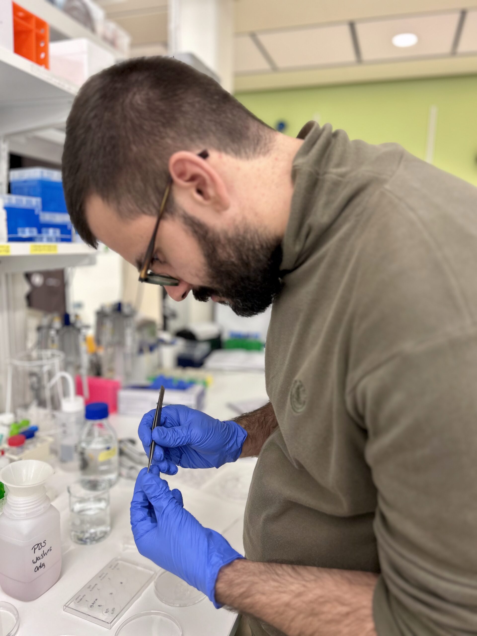
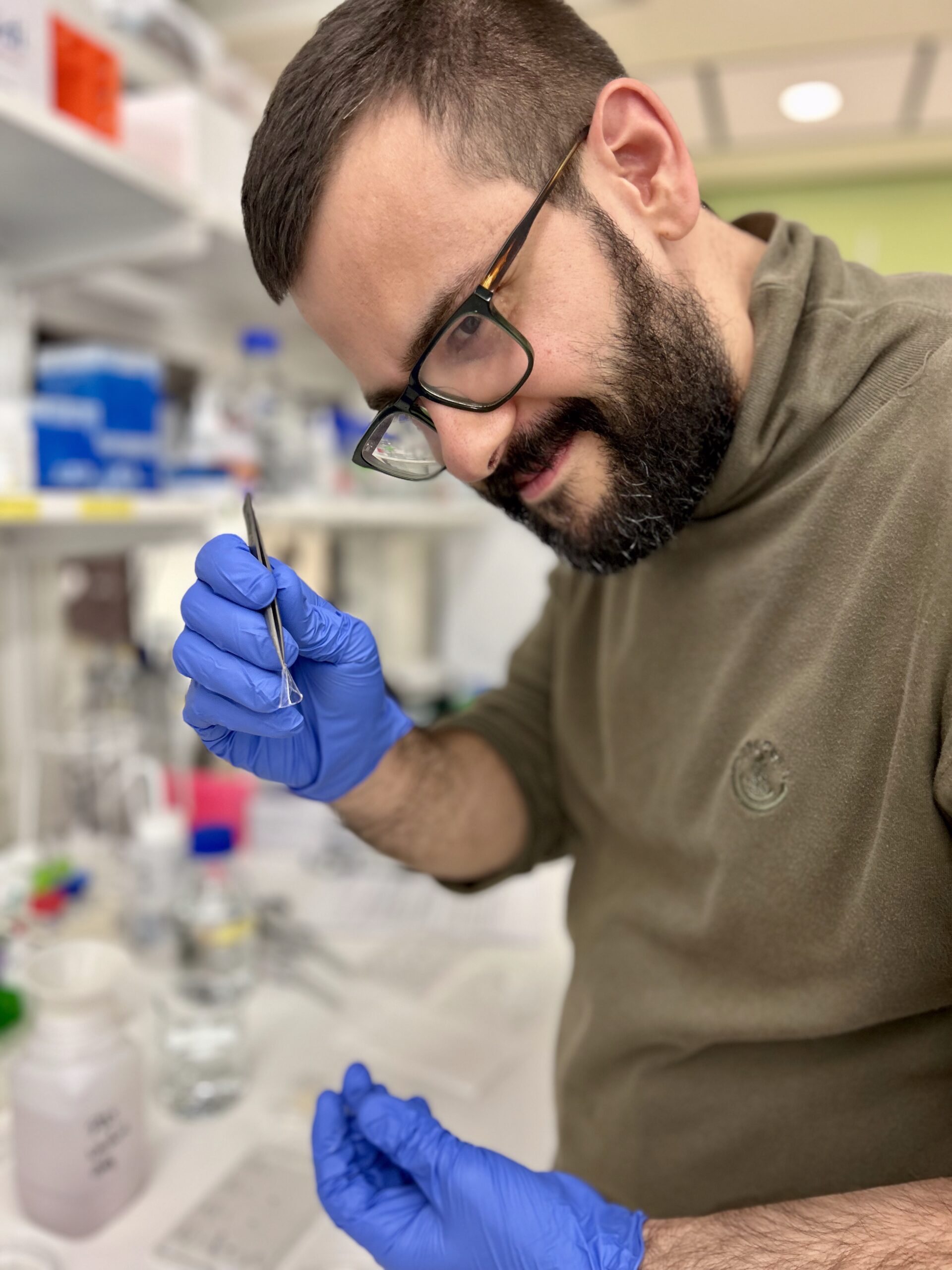
Sample preparation – The visitor tries working with the gels, based on what Ana prepared. By the end of the day, the visitor will understand how to go from a fixed mammalian cell preparation to cast and label a gel. In the meantime, Ana & her team prepare some mock gels for the visitor to handle and expand on Day 2.
Overview Imaging – The visitor assesses the correct orientation of the gels in relation to the cells, mounts the gels and acquires some initial images. They are encouraged to take the time to find the nice cell they want to take home. The trickier part is to handle and mount the gels for imaging.
Operational Imaging/Longer Imaging session – The visitor continues with imaging, taking a bit more time to find a particular cell type.
If you are interested in learning ExM from Ana and Steve, please get in touch! (ana.agostinho@scilifelab.se, steven.edwards@cilifelab.se )

The Job Shadowing programme is co-funded by the European Union as part of the EVOLVE project.
March 26, 2026
The Madrid Advanced Microscopy Center (MAdMiC) is the first Euro-BioImaging Node in Madrid (Spain). It is formed through the collaboration and close work of…
March 26, 2026
German BioImaging, within its work in the NFDI4BIOIMAGE consortium, and in collaboration with Euro-BioImaging ERIC, has launched a new survey to collect input about…
March 25, 2026
Turin, Italy – 20–22 October 2026 Early career professionals working in imaging core facilities will soon have the opportunity to strengthen essential skills beyond…