October 26, 2026
Euro-BioImaging at the EOSC Symposium
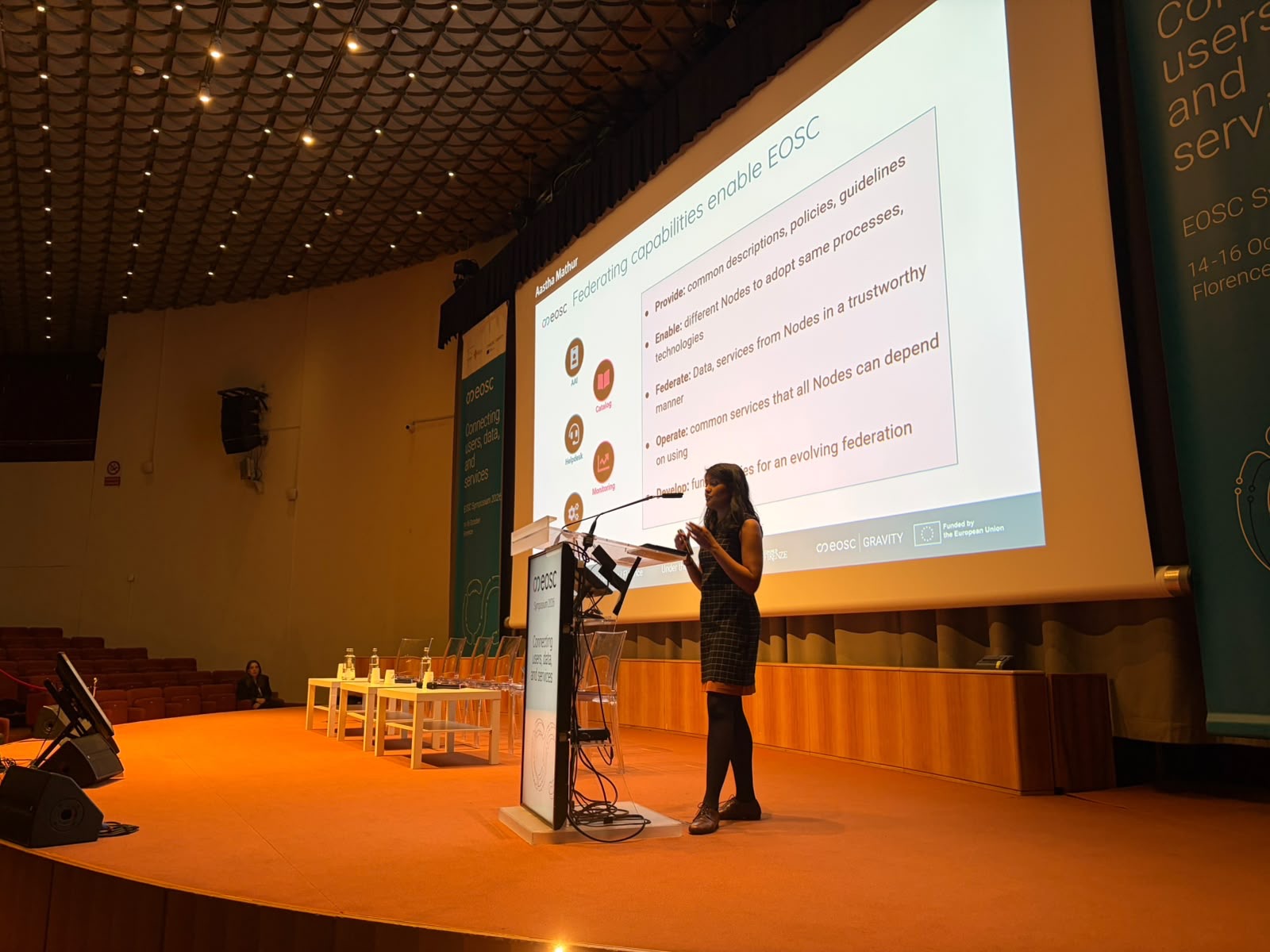
The EOSC Symposium 2026 took place from October 14-16, in Florence, Italy. More than 500 participants from 35 countries gathered there, including Euro-BioImaging’s…


As part of our series highlighting the scientists behind the Euro-BioImaging Nodes, our Scientific Ambassador, Prasanna Padmanaban met with Dr. Nina Glöckner Burmeister, Manager of the Bioimaging Core Facility at Aarhus University, part of Danish BioImaging Node. Her work brings together biological questions, imaging physics, instrument expertise and user training. At Aarhus University, Dr. Nina Glöckner Burmeister manages a busy bioimaging core facility where advanced microscopy, practical problem solving and scientific collaboration come together. The facility includes 16 microscopes and several analysis workstations. The facility has trained 400 scientists in the past two years. Its scale creates both opportunity and responsibility: researchers arrive with different samples, levels of experience and scientific timelines, while the facility team must keep complex instruments reliable and ensure that every project begins with the right imaging strategy.
Article contributed by Euro-BioImaging Scientific Ambassador Dr. Prasanna Padmanaban
Nina’s route into microscopy began at the University of Tübingen. She initially studied ecology, but a live cell imaging practical changed the direction of her career. Watching single proteins move inside living cells transformed microscopy from a laboratory technique into a way of experiencing biology as a dynamic process.
“Seeing single proteins move inside cells for the first time completely fascinated me.”
-- Dr. Nina Glöckner Burmeister
She developed her microscopy experience during her master’s degree through extensive work with confocal imaging. Her PhD then took her into fluorescence lifetime imaging, single molecule tracking and signalling research using a custom-built microscope by collaborators from the physical chemistry department. The work required her to move between biology, optics, programming and instrument development.
That interdisciplinary foundation proved ideal preparation for core facility science. Today, Nina combines scientific consultation, technology development and practical training, helping researchers translate experimental questions into imaging workflows that can produce interpretable data.
What continues to motivate Nina is the interaction between biological discovery and physical principles. Before joining Aarhus University, she worked with fluorescence correlation spectroscopy and two photon microscopy across diverse applications—from tracking beads in the trachea of mice to imaging algal fast movement and observing blood flow.
A core facility preserves that variety. One project may involve a familiar specimen and an established workflow; the next may require a new sample preparation method, an unfamiliar imaging geometry or an unconventional analysis strategy. For Nina, this combination of technologies and scientific questions is a defining attraction of the role.
“Imaging connects disciplines—and that is what inspires me.”
-- Dr. Nina Glöckner Burmeister
Managing a large facility also requires careful prioritisation. With 16 microscopes, several workstations and a large user base, requests frequently compete for time and specialist support. Each user brings a different level of experience, and each experiment has its own biological constraints.
Alongside scientific support, Nina manages the less visible infrastructure that keeps the facility functioning: budget, a grant, service contracts, repairs, administration around these activities - and long term equipment planning. The scientific and financial dimensions cannot be separated in a core facility. Reliable access depends on maintenance and investment decisions made well in advance.
Training is therefore central to the facility’s work. Researchers need more than access to sophisticated instruments; they need the practical understanding to choose appropriate settings, recognise limitations and interpret their data responsibly.
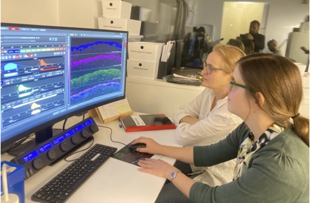
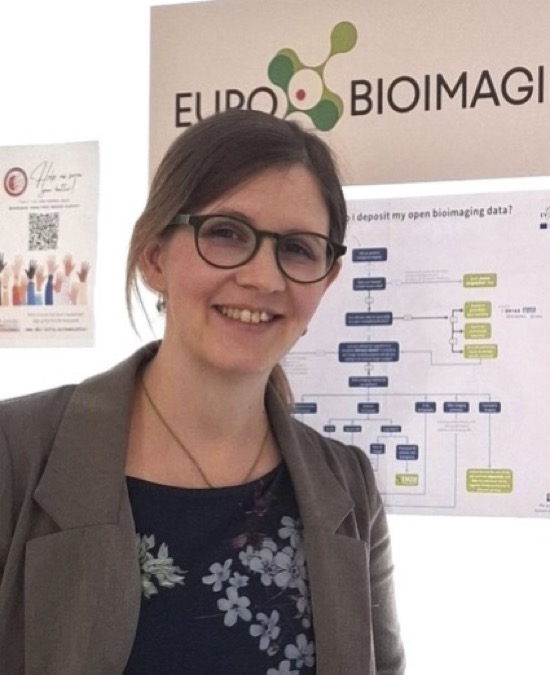
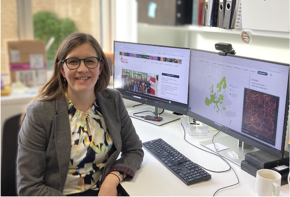
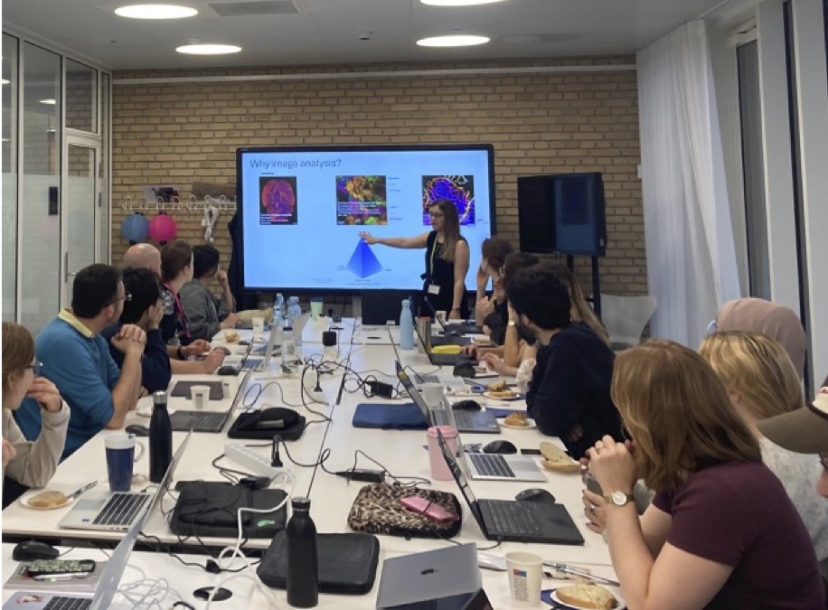
Picture 1 (left): Nina, together with co-manager Dr. Anna Lorentzen at the Stellaris FLIM confocal, Leica. Picture 2 & 3 (centre): Nina in front of the office and in the office. Picture 4 (right) Fiji image analysis course together with Image Analyses Core Facility (IACF) Copenhagen, supported by Danish Bioimaging (DBI).
Because much of the facility’s work supports biomedical research, Nina and her colleagues are expanding their capabilities in whole organ and intravital imaging. Light sheet microscopy and two photon microscopy are important parts of this development.
These approaches allow researchers to investigate living systems, larger specimens and structures located deeper within tissue. They can reveal spatial and dynamic biology that is difficult to capture using conventional microscopy, but they also demand careful attention to experimental design, sample preparation and data handling. With these complementary techniques, scientific questions can be addressed from organ-scale down to single molecule dynamics.
Nina describes the Danish imaging community as friendly, collaborative and technology driven. The Aarhus facility already works through Danish and Nordic networks, and its connection with Euro-BioImaging creates opportunities to share expertise, infrastructure and training more widely.
One example is the facility’s collaboration with SciLifeLab in Sweden on light sheet microscopy courses. These activities combine specialist knowledge with hands' on education, helping researchers understand what the technology can offer and how to design experiments that use it effectively.
“Core facilities are strongest when they collaborate. No single facility can hold every technology.”
-- Dr. Nina Glöckner Burmeister
For researchers, a connected infrastructure makes it easier to find technologies and expertise that may not be available locally. For facilities, it creates a community in which experience can be exchanged, practices improved and ambitious projects developed collectively.
For early career researchers considering core facility work, Nina emphasises the importance of a supportive and service oriented mindset. Facility scientists need to care about the success of other researchers while developing deep expertise in at least one area, such as optics, image analysis or a major imaging modality.
Technical knowledge matters, but practical judgement develops at the microscope. Real samples are rarely perfect, and many imaging problems must be solved through observation, patience and structured troubleshooting.
“Practical problem solving is the heart of imaging facility science.”
-- Dr. Nina Glöckner Burmeister
What excites Nina most about Euro-BioImaging is its ability to connect microscopists, technologies and research communities across Europe. Participation gives researchers routes to expertise and instruments beyond their home institutions, while facilities gain trusted colleagues with whom they can exchange knowledge and tackle projects that would be difficult to realise alone.
For the Bioimaging Core Facility at Aarhus University, joining this wider community is both an opportunity to contribute and a way to keep developing. Its combination of advanced technology, practical training and collaborative facility science places it firmly within a European imaging landscape built on shared expertise.
October 26, 2026
The EOSC Symposium 2026 took place from October 14-16, in Florence, Italy. More than 500 participants from 35 countries gathered there, including Euro-BioImaging’s…
October 26, 2026
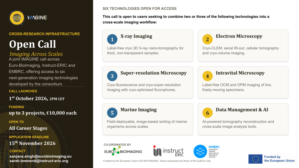
Could combining novel imaging technologies help you answer a research question—or develop a new workflow at your facility? The IMAGINE Open Call:…
October 23, 2026
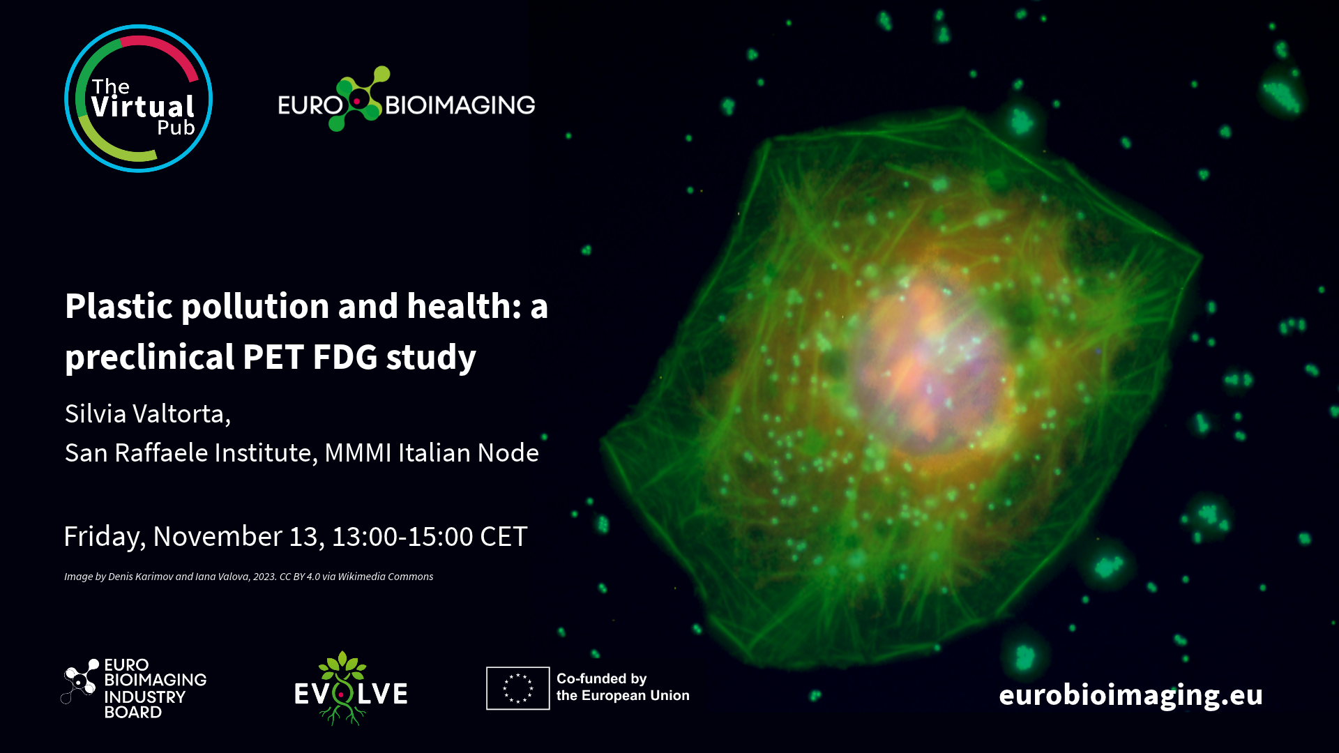
At the Special Edition Virtual Pub, “Imaging Micro- and Nanoplastics,” Euro-BioImaging users, Node experts and Industry partners will explore how advanced microscopy, clinical…