Interview with Helena Chmelová, Ph.D., from Light Microscopy Core facility at Institute of Molecular Genetics, Prague, Czech Republic, part of Euro-BioImaging’s Advanced Light And Electron Microscopy Node Prague.
Could you tell us a little about yourself?
I joined the Light Microscopy Core Facility at the Institute of Molecular Genetics of the Czech Academy of Sciences, part of Euro-BioImaging’s Prague Node, as a light microscopy specialist. I am particularly interested in imaging of living samples - cells, tissues and whole model organisms. That is why I am keen on promoting QPI microscopy technique: It represents an elegant method enabling live cell imaging over extended periods of time with low phototoxicity and good cellular contrast
Could you provide a short summary of QPI microscopy and list some applications?
Quantitative Phase Image (QPI) microscopy is a method for long-term label-free imaging of living cells. As the light passes through a transparent specimen, different parts of the specimen with different thickness and distinct refractive indices generate slight changes in the phase of the light wave. QPI uses this phase change to provide quantitative information by creating so called “phase” images where acquired phase shift values are directly proportional to cell dry mass.
This method is very useful for studying cell shape, viability, motility, polarity, growth or differentiation under various conditions. Plethora of studies focusing on drug testing, cell interactions or cancer cell biology can take advantage of this technology.
Tell us a bit more about a specific project that was done in your facility using this technology? What scientific questions were you addressing?
One of the projects which successfully exploited QPI technology in our facility focused on regulation of front-rear polarity in spreading cells. The authors wanted to evaluate the reorganization of cell mass during cell polarization, therefore, QPI was the method of choice as it enables direct quantification of cell dry mass distribution and its changes over time.
Fig: Example of quantitative phase image of a Rat2 fibroblast and its segmentation. Cell dry mass distribution is displayed as the grayscale or pseudo-coloured quantitative phase images. Author: Tomas Grousl, Laboratory of Cell Signalling, Institute of Microbiology of the Czech Academy of Sciences. Publication link: https://www.mdpi.com/2218-273X...
Why is QPI best suited to address that question?
This technology enables visualization of low-contrast cells, offers direct observation of morphological changes, including rearrangement of cell mass, and facilitates subsequent image processing and analysis.
What are some challenges of QPI? What do researchers have to pay attention to when performing these experiments?
This method is particularly convenient due to its low demands on sample preparation. As mentioned already, it makes it possible to image label-free cells without any need of contrast stains. Nevertheless, formation of the quantitative phase image can be influenced by the used media solutions and it is advisable to maintain stable conditions such as temperature, refractory index and constant volume of the solution while avoiding meniscus formation.
What other services do you provide in your facility that would be useful in combination with this type of microscopy?
At the Light Microscopy Core facility of the Institute of Molecular Genetics in Prague, we offer a wide range of light microscopy technologies from fluorescence widefield microscopy through confocal scanning or spinning disk microscopy to various super-resolution approaches. Hence, scientists here can build on their observation of cellular behaviour obtained by QPI imaging and easily dive into high-resolution techniques to further explore underlying mechanisms at subcellular or even single-molecule level.
Why should scientists choose your facility for using this technology?
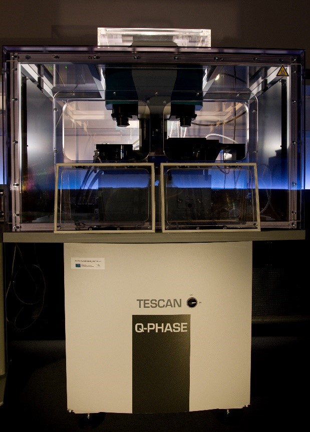
Our facility is equipped with the TESCAN Q-PHASE coherence-controlled holographic microscope for quantitative phase imaging. Strikingly, we were privileged to be present at the development of this unique patented setup as the prototype of the machine was placed and operated in our facility in between years 2014 and 2015. Since then we are in the direct contact with the developers and we have their full support for our Q-PHASE microscope and its users over and above the standard on call service, including the possibility of remote access sessions for technical maintenance. Importantly, TESCAN Q-PHASE microscope provides QPI information directly by offering automated time-lapse segmentation and cell motility measurement in very user-friendly software interface. Therefore, we believe that we can provide scientists with the comprehensive support ranging from the technical troubleshooting to the final analysis of acquired data. Additionally, TESCAN Q-PHASE microscope enables to combine QPI imaging with widefield fluorescence imaging which further broadens its versatility.
How to apply:
In July 2020, Euro-BioImaging launched a Proof-of-Concept study - in collaboration with its Nodes - making it possible to access six imaging technologies that were previously unavailable via our infrastructure. This article – the second in a series - features interviews with Euro-BioImaging Node staff, shedding light on some potential applications of these technologies - and providing advice on how to get the most out of them. We are currently accepting applications to use these technologies as part of the Proof-of-Concept study. Be part of this study and contribute to community-wide continuous technological innovation!
All scientists, regardless of their affiliation, area of expertise or field of activity can benefit from Euro-BioImaging’s pan-European open access services.Potential users of these new technologies are encouraged to submit project proposals via our website. To do so, you can Login to access our application platform, choose the technology you want to use and the facility you wish to visit, then submit your proposal. All applications will be processed by the Euro-BioImaging Hub. As usual, users will benefit from advice and guidance by technical experts working at the Nodes, training opportunities, and data management services.
A Multiplexed Vision: Highlights from the Special Edition Virtual Pub on Spatial Transcriptomics
On June 12th, Euro-BioImaging hosted its latest Special Edition Virtual Pub, bringing together over 200 researchers and technology developers working at the cutting…
Cellular Imaging Hungary Node expands expertise in advanced neurophotonics
The Euro-BioImaging Cellular Imaging Hungary Node has expanded its service portfolio with the addition of the BrainVisionCenter (BVC) in Budapest. Following a successful…
The UK Euro-BioImaging Node expands from seven to thirteen sites!
We’re delighted to announce that the UK Euro-BioImaging Node is expanding, growing from seven to thirteen sites following a successful upgrade application and…