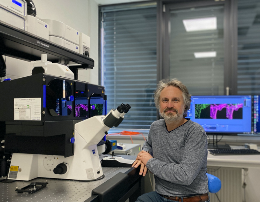
The Advanced Light Microscopy and Medical Imaging Node in Brno, Czech Republic, has just invested in a new, super-resolution microscope that can perform Structured Illumination Microscopy (SIM) and Single Molecule Localisation Microscopy (SMLM). In this interview, Milan Esner, Deputy Head of the Brno Node, explains the range of projects that will be supported with this technology and the scientific questions that can be addressed. Great news, Euro-BioImaging users are eligible to use this state-of-the-art new technology in Brno!
And for imaging projects carried out between July 2020 and June 2021, apply for funding to cover access costs from Czech BioImaging. Deadline: April 18, 2021.
Tell us who you are, where your facility is based, and what your role is.
My name is Milan Ešner, and I am the Head of CELLIM (Cellular Imaging Core Facility), a light microscopy core facility of CEITEC-MU. CELLIM was created in 2016 and since then has continuously grown – in terms of the number of operating instruments and also the number of users. Despite Covid-19 restrictions last year we served more than 100 users in total. CEITEC-MU is located within the university campus of Masaryk University in Brno, the second largest city of the Czech Republic. Due to our location on the university campus, we have many users from neighboring faculties, mostly from the Faculty of Medicine and the Faculty of Life Sciences. Therefore, we have a wide-range of imaging requirements, from smallest unicellular organisms, mammalian cells, tissue organoids to whole organisms such as mouse embryos or plants. These requirements are reflected by the diversity of our instruments – from simple stereomicroscopes, larger zoom microscopes, wide-field systems to advanced imaging confocal systems and the latest super-resolution microscope.
An important aspect of our work, besides instrument maintenance is user support – training, assistance and courses. Most of our training is with individuals face-to-face, to assure the maximum quality of training. We are also providing expertise training and access to image analysis – such as deconvolution and object detection and quantification. Every year we also organize several workshops on light microscopy. These courses are open for external users and are announced via the Euro-BioImaging portal. Unfortunately, due to COVID-19 related restrictions, most of our courses were shifted to virtual format in 2020-2021.
Your facility has just invested in a new super-resolution microscope, which can perform SIM and Single Molecule Localisation Microscopy (SMLM). Please tell us a bit more about the capabilities of this system.
We purchased the SIM/SMLM system from Carl Zeiss, Elyra 7 – Lattice SIM. This instrument has several modalities, like structural illumination microscopy (SIM), total internal reflection microscopy (TIRF) and single molecule localization microscopy (SMLM). As mentioned above we have many diverse biological samples, which is why we decided to purchase a microscope with SIM capabilities. Despite the fact that the resolution with SIM is not quite as good as SMLM or STED, it is very universal and the main advantage is that it can be used for the same samples as for confocal microscopy. With SMLM you obtain much higher resolution, but you have to optimize your staining protocol and subsequent analysis.
Why did you decide to invest in this machine?
Super-resolution imaging/nanoscopy was missing in our facility and such modality was not accessible anywhere on the whole university campus or Brno region. Thanks to CzechBioimaging’s national programme for financing research infrastructures we obtained finances to purchase such a system for our facility. As we are a facility serving many research groups with diverse thematics, we aimed to purchase the most universal and versatile system possible, which could be used for many different biological models and for fixed and living specimens. For us, the SIM (Structured Illumination Microscopy) system is just that. At the time we made our decision, Zeiss had just released this revolutionary Lattice SIM, using a lattice pattern instead of classical line grid structures. The choice of single molecule localization microscopy (STORM/PALM) is connected to this decision, as the Zeiss SIM system comes together with SMLM.
What kind of projects do you hope to support using this technology? What scientific questions will you be able to address?
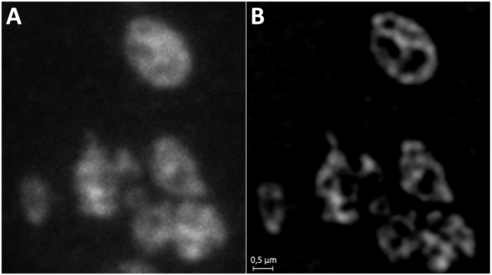
Already several users from different research groups were succesfully using the system. For the moment, the system has been used mainly for adherent mammalian cells. In the example below, you can see stained mitochondria. A) sample imaged in wide-field, B) the same sample but imaged with SIM.
What other services do you provide in your facility that would be useful in combination with this type of microscopy?
We also provide image/data analysis support to our users. We are using various open source and commercial image analysis tools running on our computers. Those can be operated remotely – meaning the user does not need to come to our facility. This is quite good given current travel restrictions. For image analysis, we also have a close collaboration with Prof. Michal Kozubek from Centre of Biomedical Image Analysis department of Faculty of Informatics, Masaryk University.
Why should scientists choose your facility for using this technology?
This system is new and is not accessible in many different locations for the moment. Compared to classical SIM, the new Lattice SIM is much faster and more robust. And, researchers interested in this technology and other technologies available at our facility can apply for open access via Euro-BioImaging.
How can users fund a visit to your Node?
Twice a year, Czech BioImaging offers the possibility to apply for small grants to cover expenses for measurements – instrument time and other access costs. More information can be found at:
Global BioImaging Advance the Conversation on Sustainable Biomedical Imaging Facilities
How can biomedical imaging facilities remain scientifically excellent while becoming more financially, environmentally, and operationally sustainable? These questions were at the heart of…
Moara Lemos: New insights from Global BioImaging’s Job Shadowing programme
Moara Lemos is a Brazilian researcher and imaging scientist. Her award winning research pushes the boundaries of structural biology, using advanced cryo-electron microscopy…
A Multiplexed Vision: Highlights from the Special Edition Virtual Pub on Spatial Transcriptomics
On June 12th, Euro-BioImaging hosted its latest Special Edition Virtual Pub, bringing together over 200 researchers and technology developers working at the cutting…