
April 13, 2026
We are hiring a Data Architect/Knowledge Engineer
Euro-BioImaging Bio-Hub team, hosted by EMBL in Heidelberg, is hiring a Data Architect/Knowledge Engineer – Euro-BioImaging (AI4Access) to…

Today is the International Day of Women and Girls in Science. And tomorrow. And the day after that. The images below show our everyday. Women working in science across our research infrastructure. Thanks to all of the colleagues, Node staff and Euro-BioImaging users who are featured. Please read below to learn more about their work.

Maria Karolina Andersen, post-doc researcher at the Norwegian Institute of Science & Technology (NTNU) - Trondheim
Maria Karolina Andersen studies prostate cancer, as part of the ERC-funded ProstOmics project. To further explore the role of cholesterol in prostate cancer, she applied for access to Euro-BioImaging’s Facility of Multi Modal Imaging AMMI Maastricht Node, where a highly sensitive MALDI2 MSI instrument is available. Read the article
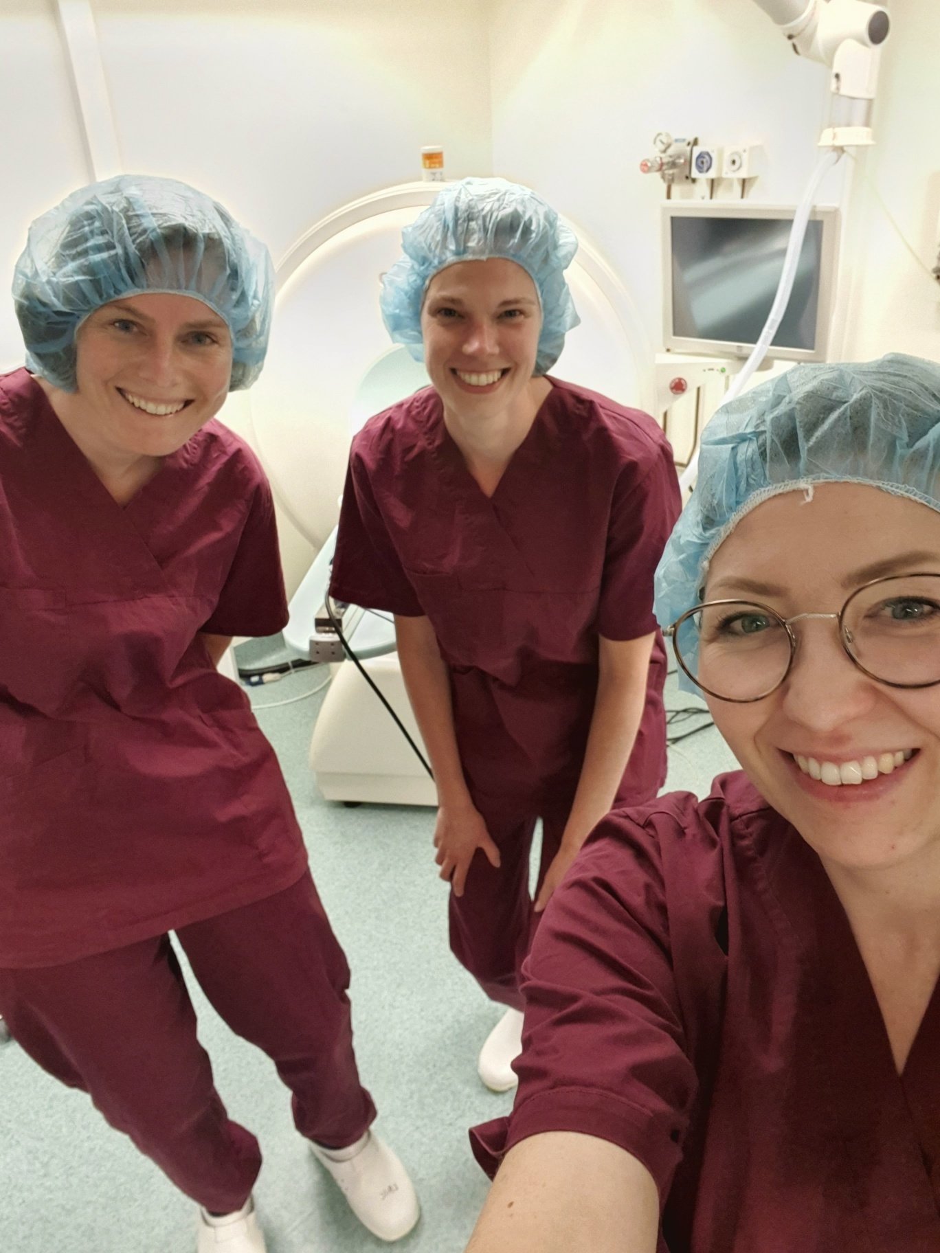
Irma Mahmutovic Persson, staff researcher at Lund BioImaging Centre (LBIC), Lund University, part of the Euro-BioImaging Node Swedish National Microscopy Infrastructure (NMI)
Irma splits her time between the 9.4T preclinical MRI platform and the nuclear medicine platform, working with SPECT-PET-CT scanners. She contributes to study design, logistical planning, animal handling, and data acquisition. In some projects, she supports researchers with specific technical procedures such as radiotracer injections, while in others she is involved from hypothesis development to final data analysis. Learn more about her work in this interview
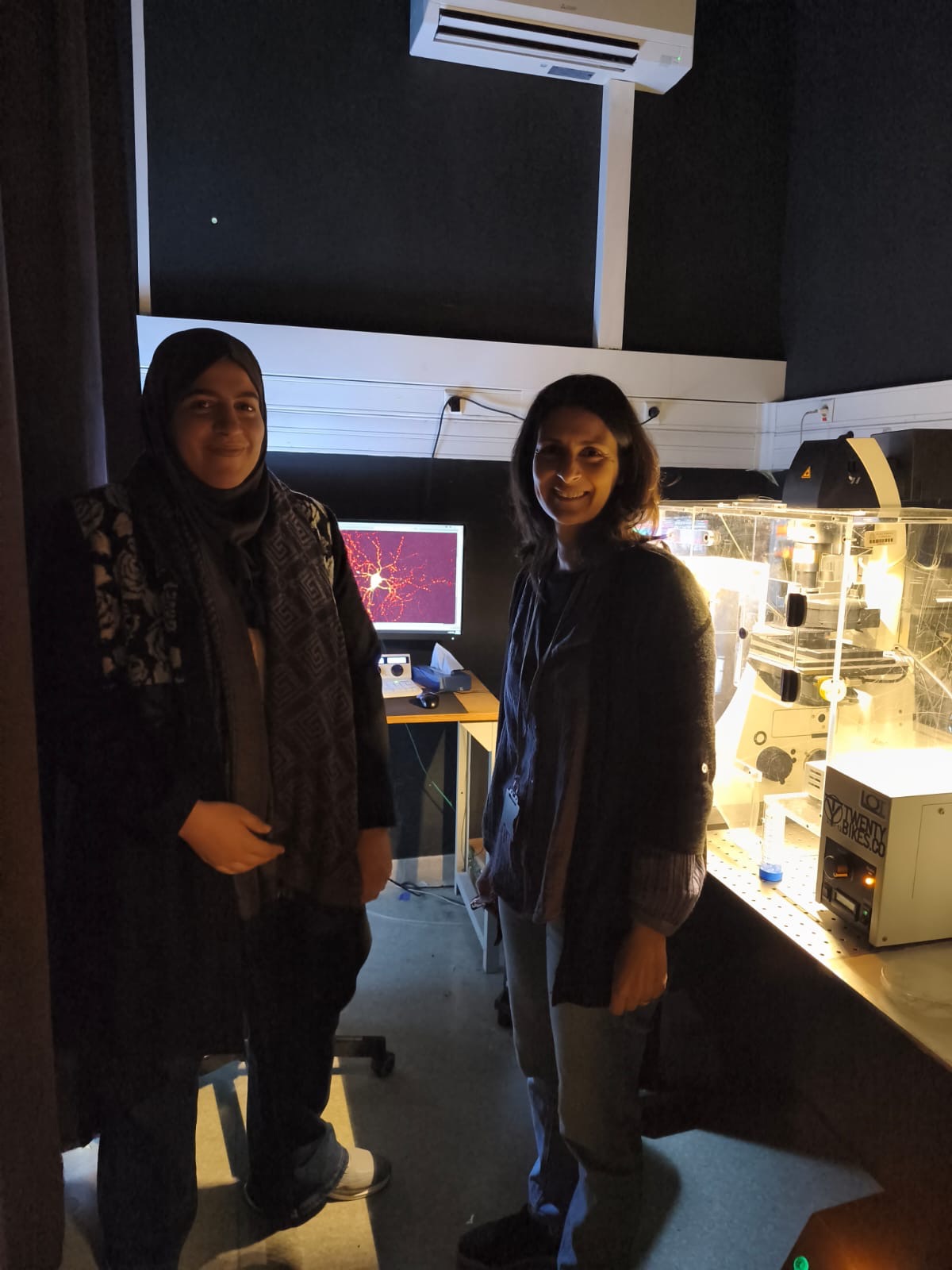
Samira Benadaa, Imachem ENS, part of France-BioImaging, and Wiame Aissoug, CRBT Constantine, Algeria
As part of the Imaging 4 All (i4A) Pro Track funding programme, Wiame AISSOUG spent several months with Samira Benadda at Imachem ENS, where she received training on management of imaging facilities as well as hands on training on specialised microscopy (Expansion Microscopy, TIRF, confocal, spinning disk, Live cell imaging), image analysis, as well as sample preparation. More information
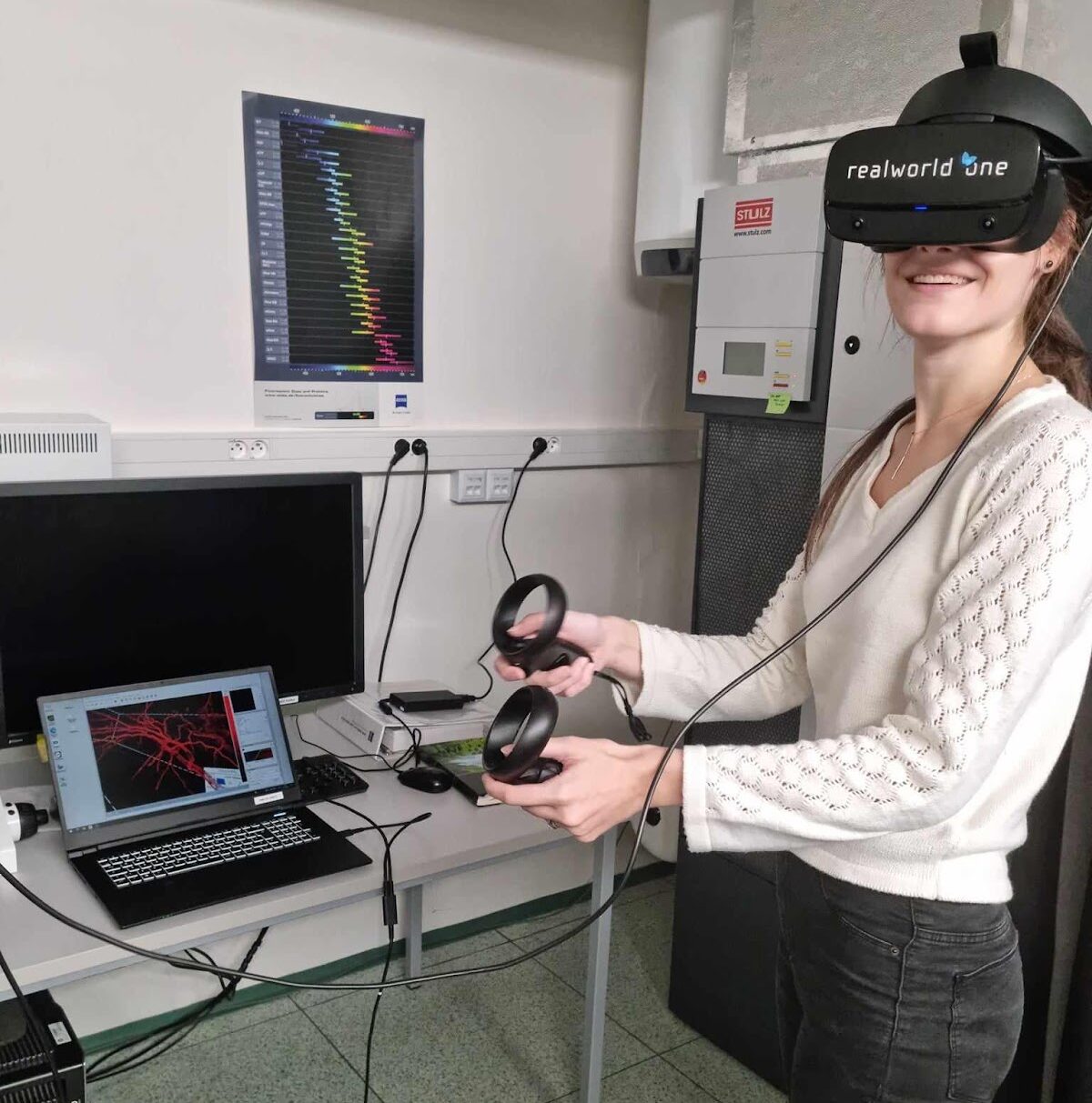
Katerina Kaduchova, PhD student Centre of Plant Structural & Functional Genomics, Institute of Experimental Biology, Czech Academy of Sciences, Olomouc
In October 2023, Katerina attended a two-day course on “Multi-modal light microscopy imaging in plant research,” organized by Katerina Malinska, IFIEB in Prague, part of Euro-Bioimaging’s Advanced Light & Electron Microscopy Prague Node. During this workshop, she tried out several microscopy systems, experimented with root/growth tracking software, and discovered how Virtual Reality can contribute to image analysis. Read about it Picture taken by: Beáta Strejčková (shared by @pecinka_grp on X)
Jessica Aye Valdivia Pérez from Centro de Investigaciones en Química Biológica de Córdoba (CIQUIBIC), Argentina
Jessica's research (funded by CONICET) focuses on how amphiphilic drugs interact with lipid membranes using advanced fluorescence techniques to uncover their biophysical mechanisms of action and guide future therapeutic design. Her visit to the Advanced Optical Microscopy Centre (AOMC) at University of Hasselt, part of Euro-BioImaging’s Flanders BioImaging Node, where she performed a series of FLIM experiments, was supported by Global BioImaging’s Imaging 4 All initiative. Learn more Photo courtesy of the University of Hasselt.
Moona Huttuunen, Senior Lecturer, University of Jyväskylä, ISIDORe User at the FiaM Node
Moona Huttuunen received ISIDORe funding to study Poxvirus-induced extracellular vesicles in infection and cell biology with Light Microscopy and Super Resolution imaging at the Finnish Advanced Microscopy Node, work she presented at the Special Edition Virtual Pub: “Showcasing Research & Stories from ISIDORe Users”


Tiina Saanijoki (right), Coordinator of the Finnish Biomedical Imaging Node
Tiina coordinates access to the FiBI Node and supports discoveries that have an impact on Finnish healthcare services, and services used in clinical diagnostics. Work at the FiBI Node of Euro-BioImaging has also influenced international health care standards. Learn more
Estefanía Calvo Alvarez, a researcher at the Department of Pharmacological and Biomolecular Sciences, University of Milan
Estefanía Calvo Alvarez, a researcher at the Department of Supported by ISIDORe funding, Estefanía Calvo Alvarez used remote multimodal imaging services from Euro-BioImaging’s FiBI Node to analyse, for the first time, Leishmania tarentolae parasites (Ltar)’s homing, tissue tropism, and innate inflammatory responses in both immunocompetent and immunosuppressed mice. Discover her work
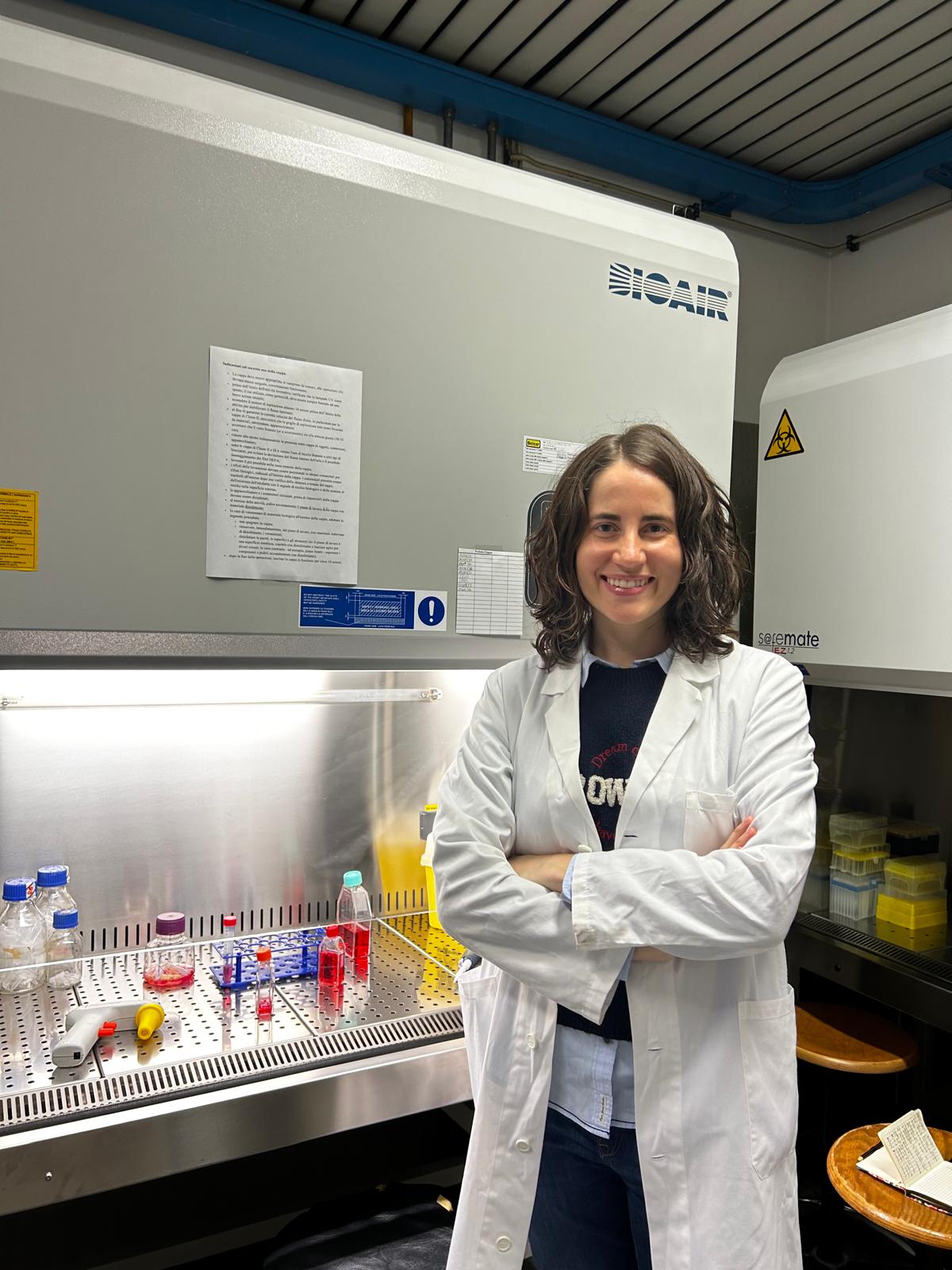

Palina Nepachalovich, PhD student, Center of Membrane Biochemistry and Lipid Research at TU Dresden
Palina had never used a microscope when she applied for access at Euro-BioImaging’s Sofia Node in December 2021, as part of the Euro-BioImaging pilot User Access fund. Her promising project proposal, involving “Membrane disruptions and repair in cell death processes,” was selected for funding. Learn more about her experience and why this visit is so important to her research – and her career.
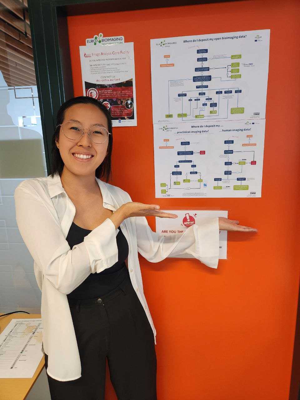
Tricia Loo, Image Analyst, Image Analysis Core Facility, Danish BioImaging Node
From providing open services in image analysis to life scientists and medical researchers, to organising training classes, Tricia Loo helps researchers visualize, analyze, and extract quantitative information out of their bioimaging datasets and much more! Here she is pictured with the Image Data Repository Decision Tree.
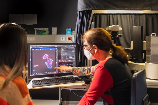
Beate Neumann (right), Senior Scientific Officer, ALMF, EMBL Node
Beate Neumann plays an advisory role with researchers who visit the EMBL Node, helping them to set-up experiments to get the results they are looking for and training them with the techniques. Here she supports Chiara Cassiolli's project to better understand SupraMolecular Attack Particles (SMAP), with a high-throughput RNAi screen aimed at elucidating how the pathways that regulate the trafficking of individual SMAP components converge to allow SMAP biogenesis. Learn more Photo Courtesy of EMBL/PhotoLab/Massimo del Prete

Marja Lohelo, Intravital imaging specialist, Biomedicum Imaging Unit, University of Helsinki, part of Finnish Biomedical Imaging Node
Marja Lohela was recently involved with a remote access project funded by ISIDORe. In the article below, she shares her experience, providing insights into - and solutions to - some of the challenges of running a remote user project. Read her perspectives
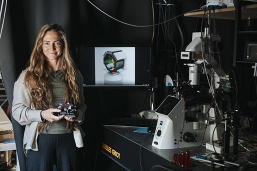
Dr. Mia Kvåle Løvmo, Institute of Biomedical Physics, Medical University of Innsbruck
Dr. Mia Kvåle Løvmo and Prof. Monika Ritsch-Marte, together with their collaborators at the Medical University of Vienna, part of Austrian BioImaging, have launched an exciting project to combine for the first time acoustic trapping and rotation with OCT. Read the full story

Claudia Pfander, Euro-BioImaging Hub Team & Yara Reis, Global BioImaging
Connecting with the imaging community both in Europe and globally - is important part of our mission. At ELMI 2025, we met an amazing number of researchers, core facility staff and industry partners at our booth. Here, Claudia Pfander, Euro-BioImaging Industry Board coordinator is pictured with Yara Reis of Global BioImaging. Learn about ELMI 2025
April 13, 2026
Euro-BioImaging Bio-Hub team, hosted by EMBL in Heidelberg, is hiring a Data Architect/Knowledge Engineer – Euro-BioImaging (AI4Access) to…
April 13, 2026
As part of our ongoing series spotlighting the scientists working at imaging facilities across the Euro-BioImaging realm, we sat down with Przemek Krawczyk, a…

April 9, 2026
Euro-BioImaging ERIC is deeply grateful to announce that the European Union has entrusted our infrastructure with funding to shape the future of imaging and…