
March 9, 2026
Innovation and collaboration at Euro-BioImaging’s AMMI Maastricht Node
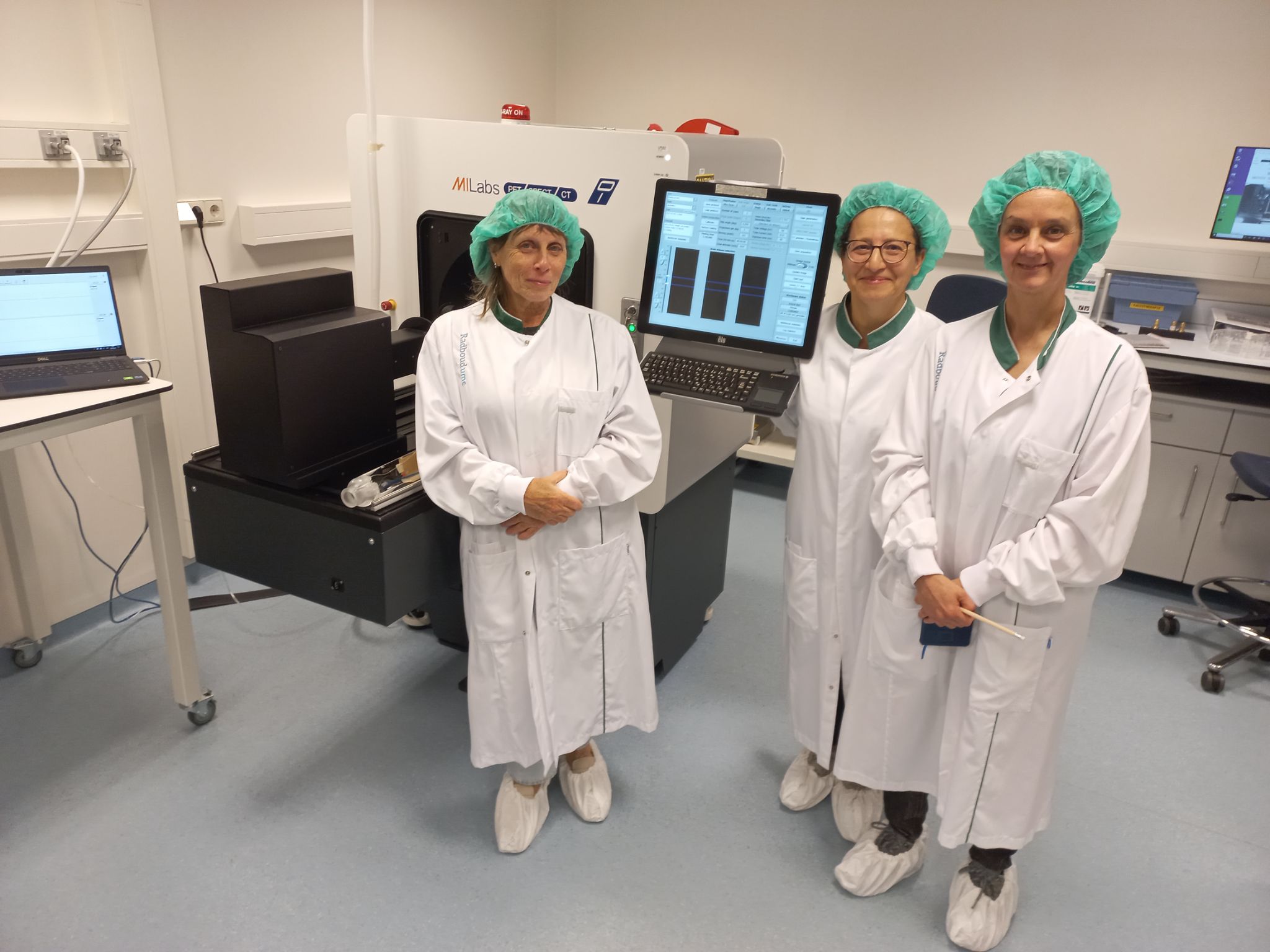
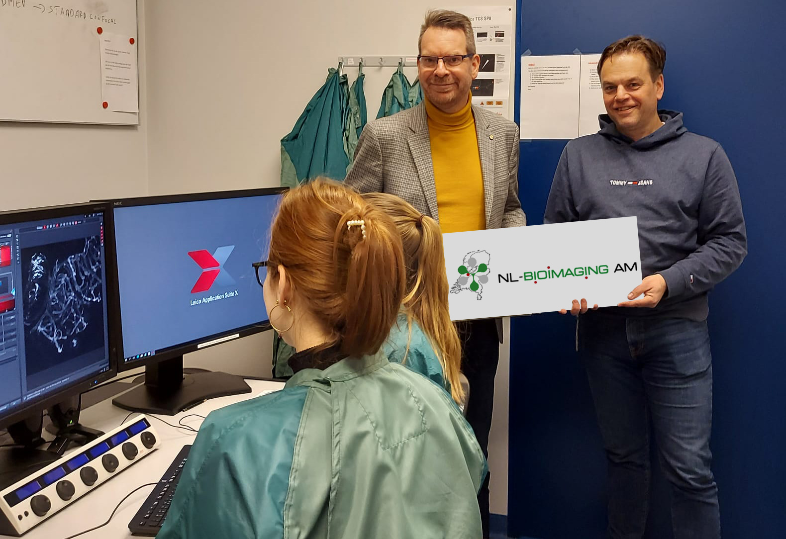
Euro-BioImaging’s Advanced Microscopy and Molecular Imaging (AMMI) Node is a single-sited mixed Node centrally located in Maastricht, in…
The Kingdom of the Netherlands, a founding member of Euro-BioImaging, proudly hosts ten nodes across the country:
Learn more about the Nodes and get an overview of news and interactions with the Netherlands below.

3
Facilities
N/A
External Users

N/A
User Publications
N/A
Remote Users

N/A
Training courses

N/A
Staff involved
5
Facilities
10
External Users
50
User Publications
0
Remote Users
2
Training courses
5
Staff involved
1
Facilities
7
External Users
57
User Publications
0
Remote Users
5
Training courses
8
Staff involved
3
Facilities
35
External Users
7
User Publications
20
Remote Users
3
Training courses
12
Staff involved
2
Facilities
14
External Users
~20
User Publications
1
Remote Users
4
Training courses
8.4
Staff involved
8
Facilities
5
External Users
18
User Publications
0
Remote Users
2
Training courses
7
Staff involved
1
Facilities
183
External Users
5
User Publications
N/A
Remote Users
5
Training courses
2.5
Staff involved
3
Facilities
9
External Users
~40
User Publications
0
Remote Users
6
Training courses
9
Staff involved
2
Facilities
19
External Users
~40
User Publications
0
Remote Users
7
Training courses
3.1
Staff involved
March 9, 2026
Euro-BioImaging’s Advanced Microscopy and Molecular Imaging (AMMI) Node is a single-sited mixed Node centrally located in Maastricht, in…

January 13, 2026
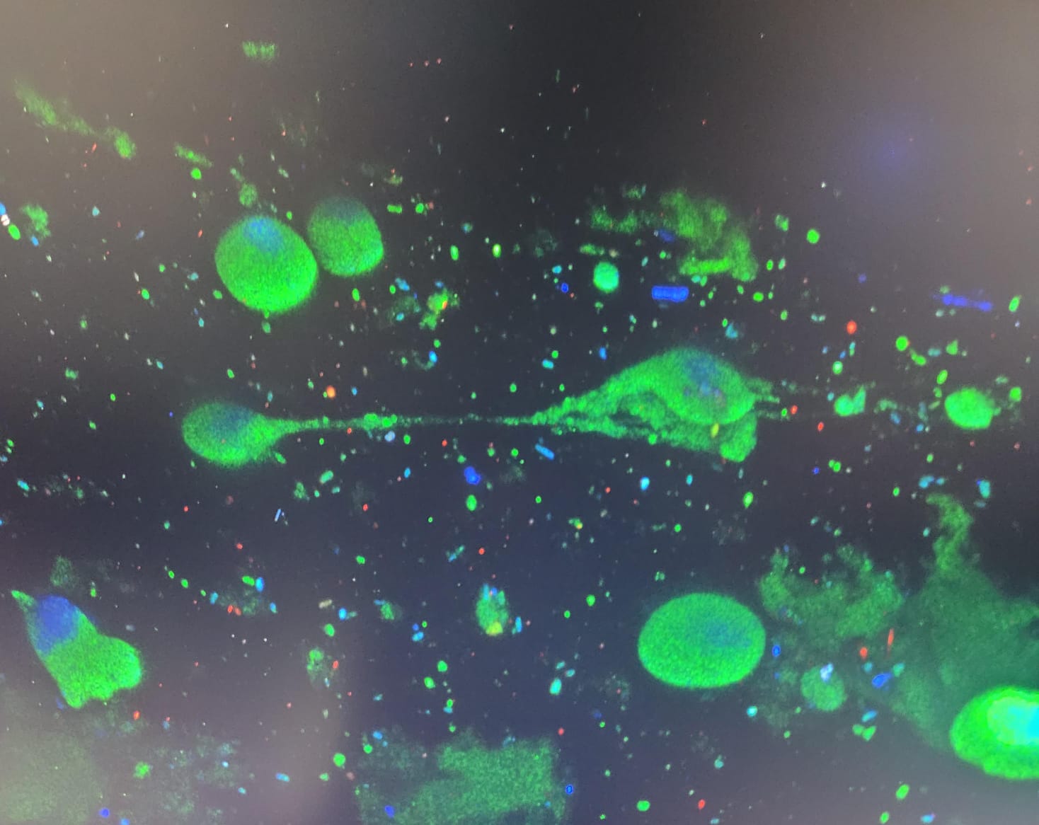
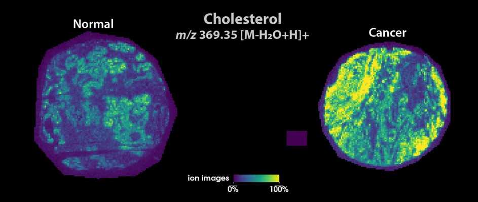
Through the canSERV project, Euro-BioImaging Nodes continue to support innovative cancer research across Europe. One recently completed user project, carried…
December 2, 2025
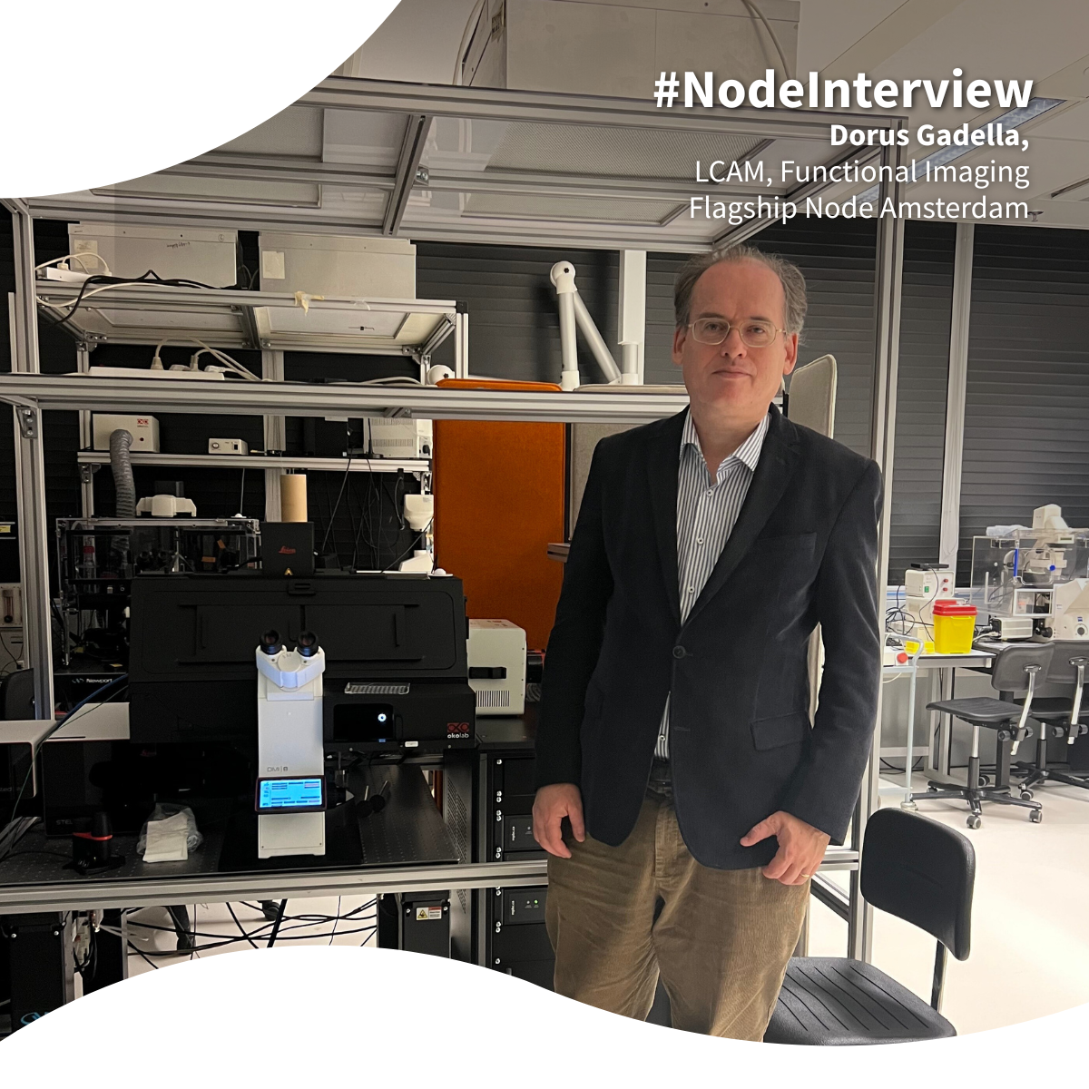
As part of our ongoing Node interview series led by Scientific Ambassadors, Vanessa Coelho-Santos, Group Leader at University of Coimbra, sat down with Professor…

November 25, 2025
We are pleased to announce that the Euro-BioImaging family is growing – and consolidating! On November 19, 2025, three new Nodes joined our infrastructure,…

October 10, 2025
We will host the Special Edition Virtual Pub “Imaging in Food Science” on Friday, October 17 from 1-3 pm CEST. It will focus on the…

October 10, 2025
We will host the Special Edition Virtual Pub “Imaging in Food Science” on Friday, October 17 from 1-3 pm CEST. It will focus on the…
August 11, 2025
Euro-BioImaging’s Population Imaging Flagship Node is embedded within the department of Radiology & Nuclear Medicine at the Erasmus…

July 31, 2025
The first-ever Euro-BioImaging Image Data Community Days, held online from April 7–11, 2025, concluded with resounding success, providing a dynamic platform for participants to…
July 21, 2025
Would learning to use the most cutting-edge imaging instruments help you answer your scientific question? Could world-class technology accelerate your research? We spoke to…
July 15, 2025
As part of the EVOLVE Mentoring Masterclass series, Euro-BioImaging was proud to host Dr. Sylvia E. Le Dévédec on June 23, 2025, to share…
June 30, 2025
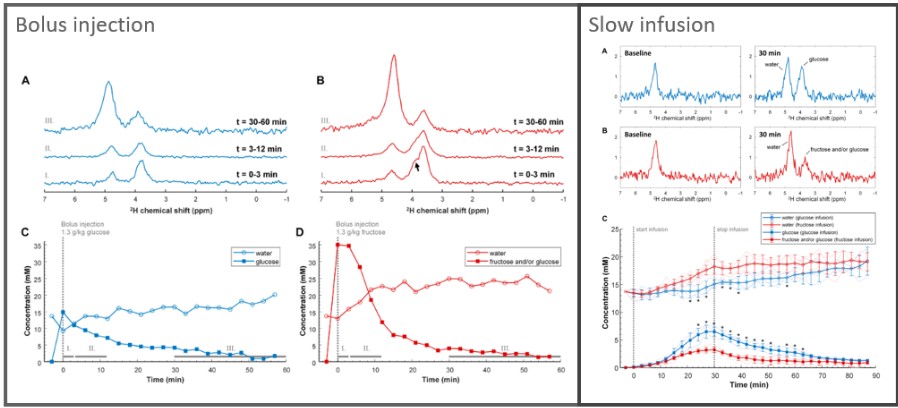
Chronic intake of high amounts of fructose has been linked to the development of metabolic disorders caused by the almost complete clearance of fructose…
June 30, 2025
With the occasion of the Board meeting which took place in Amsterdam last May, the Med-Hub section Director Linda Chaabane and the Med-Hub Head…
May 27, 2025
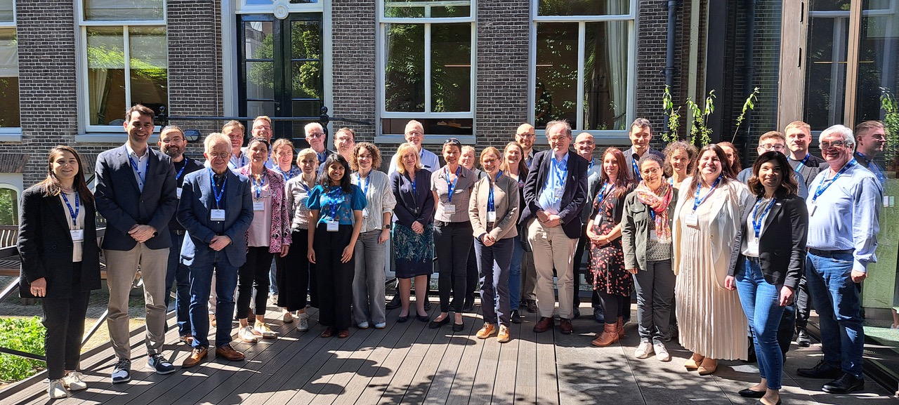
The 12th Euro-BioImaging Board meeting took place in Amsterdam, from May 13-14, 2025, hosted by our colleagues from NL-BioImaging. It was the occasion for…
March 12, 2025
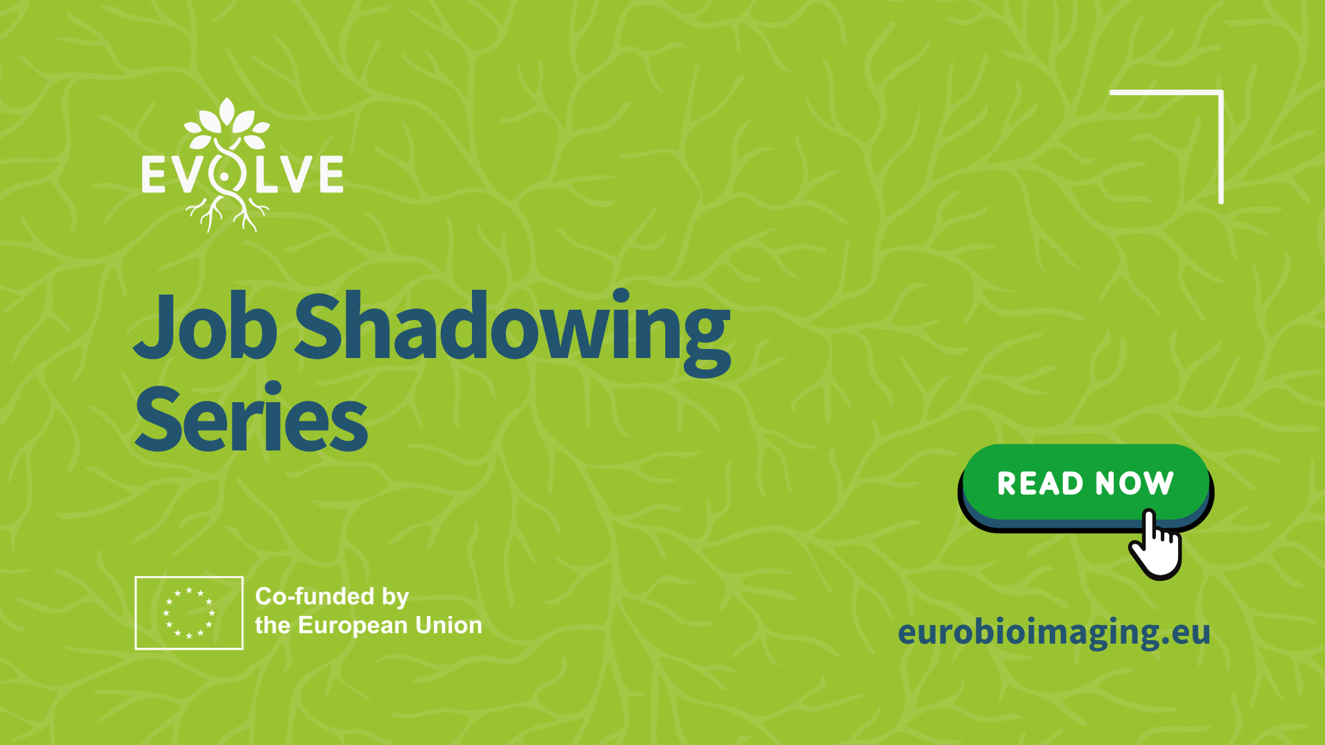
The EVOLVE Job Shadowing program is a fantastic opportunity for Node staff to immerse in the daily life and operations of other Nodes of…
February 12, 2025
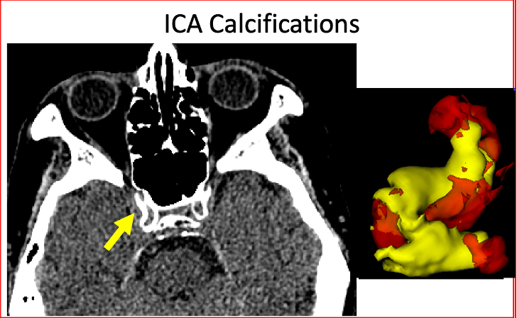
What could be more fascinating than to explore the prevalence of cardiovascular disease in populations as diverse as ancient mummies, modern Americans, and people…

October 21, 2024
Euro-BioImaging is thrilled to be part of ILLUMINATE (Increasing Lutetium production while leveraging metabolic imaging to enhance theranostics effectiveness), a pioneering 54-month public-private partnership…

June 20, 2024
The 2024 Annual Congress of the European Association for Cancer Research (EACR 2024) took place in Rotterdam from June 10 to 13, 2024. The…

July 23, 2023
What is Euro-BioImaging? What is open access to imaging? As part of a series spotlighting Euro-BioImaging Nodes, we asked Marc van Zandvoort, Chair of…

June 19, 2023
This year, the European Light Microscopy Initiative meeting took place in Noodwijkerhout, the Netherlands, organised by NL-BioImaging. Several Hub team members were able to…
February 20, 2023
Three hundred years after Antoni van Leeuwenhoek, father of live-cell microscopy, performed his last amazing research, a 25 Million Euro grant from the…
September 1, 2022
How does prostate cancer become aggressive and life-threating? That is a question that Maria K. Andersen, post-doc researcher at the…

July 19, 2022
One of Euro-BioImaging’s key missions is to facilitate excellent science by providing open access to imaging technologies and expertise. Users come from many backgrounds…

May 24, 2022
The Challenges Framework flagship Node organizes a series of Grand Challenges in Medical Image Analysis (www.grand-challenge.org), focused at standardized evaluation…
March 21, 2022
In 2021, Euro-BioImaging launched its pilot call for funded user projects to be supported by an internal Euro-BioImaging User Access Fund.

January 18, 2021
Imaging technologies can help scientists understand how plant tissues respond to stress, such as drought, heat, or other environmental factors. Characterising the mechanical properties…