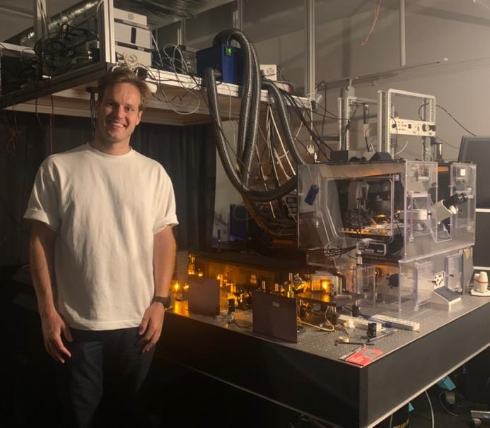
Correlative Light and Electron Microscopy of Plant tissues
Imaging technologies support research into the structure and function of plants, shed light on plant health, resilience and adaptability, and help answer agroecology-related research…
March 23, 2023
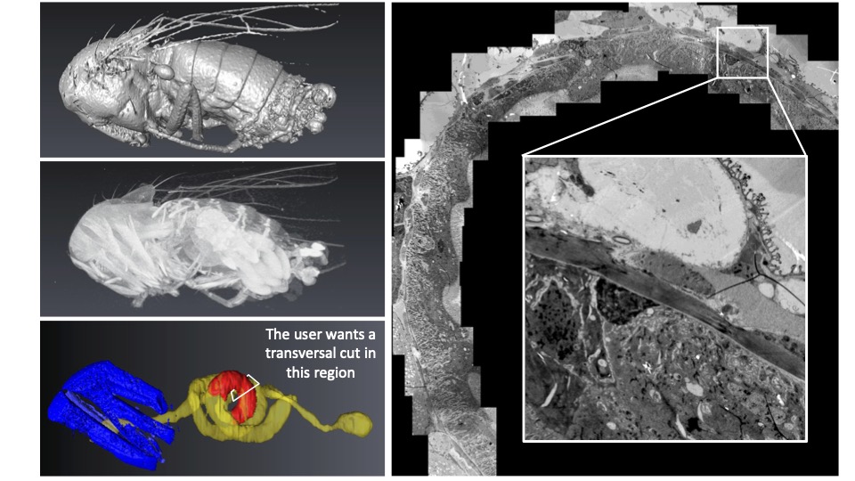
CXEM: Finding a needle in a haystack
Correlative X-ray imaging and electron microscopy (CXEM) is the combination of X-ray imaging and electron microscopy. It is a correlative approach that makes it…
March 8, 2023
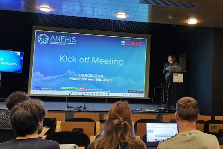
ANERIS Project: Towards Operational Marine Biology
Euro-BioImaging is delighted to be part of the ANERIS (operAtional seNsing lifE technologies for maRIne ecosystemS) project, which kicks off this week in Barcelona. The…
March 7, 2023
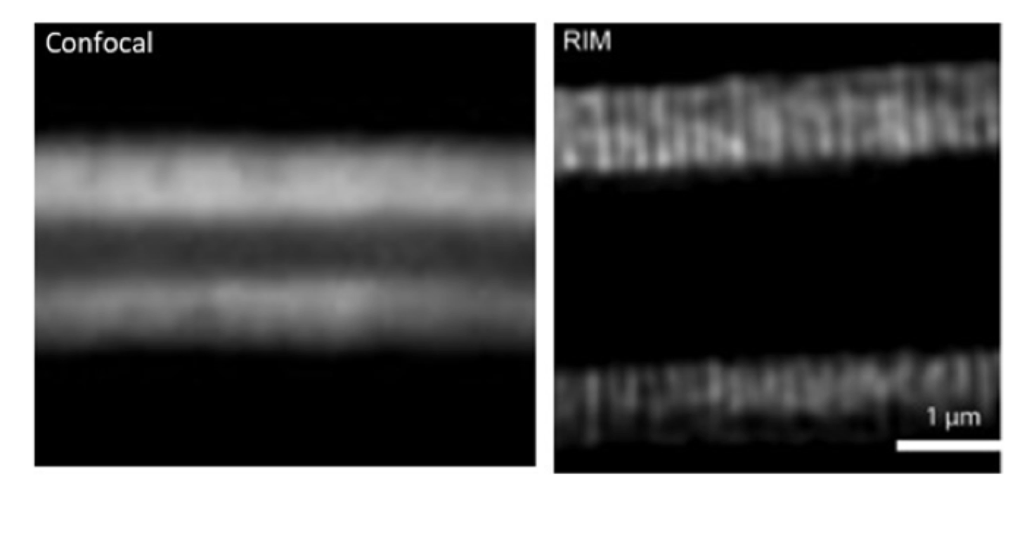
A powerful high speed, low phototoxicity microscopy method to achieve super-resolved images
Are you interested in looking at tissues or other thick samples in high resolution? We spoke to Marc Tramier, a group leader at the…
December 12, 2022
Image data analysis services at France BioImaging
Today, imaging scientists are producing datasets of increasing volume, complexity, and information content. To realize the full potential of imaging data, Euro-BioImaging Nodes offer…
November 15, 2022
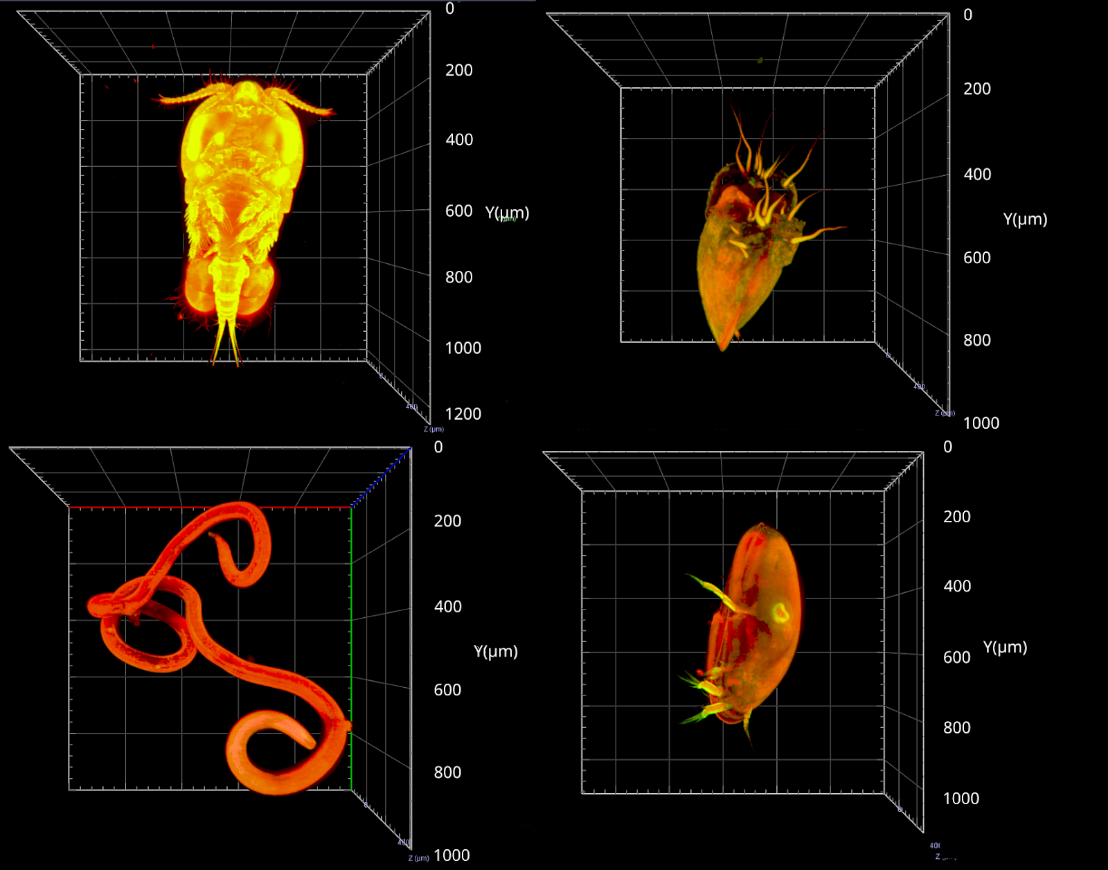
Meiofauna – the ocean’s next frontier
Meiofauna are tiny marine organisms that range in size from 20 microns to 1 milimetre. They are present everywhere in the sea, from the…
June 29, 2022
Using super resolution live cell imaging to understand cell death during stroke
Scientists have made extraordinary progress in understanding stroke and the effects it has in the brain, but are still exploring the finer details of…
March 15, 2022
Studying AMPA Receptor Dynamics with Lattice Light Sheet and Multiphoton Microscopy
Euro-BioImaging is organizing an online User Forum on April 5, 2022, from 14:00-17:00 CEST. This event will highlight the importance of…
February 17, 2022
User Access to France-BioImaging supported by CNRS grant
The Institut des Sciences Biologiques (INSB) of the French Centre National de la Recherche Scientifique (CNRS) has launched a call for projects to…
May 27, 2020
Imaging technologies used to understand COVID-19 infection
Euro-BioImaging’s French Node in Bordeaux is contributing to an important study led by the University of Bordeaux to understand COVID-19 infection and inflammatory response…